Overview
Hover to read
Click to keep this side open
Overview
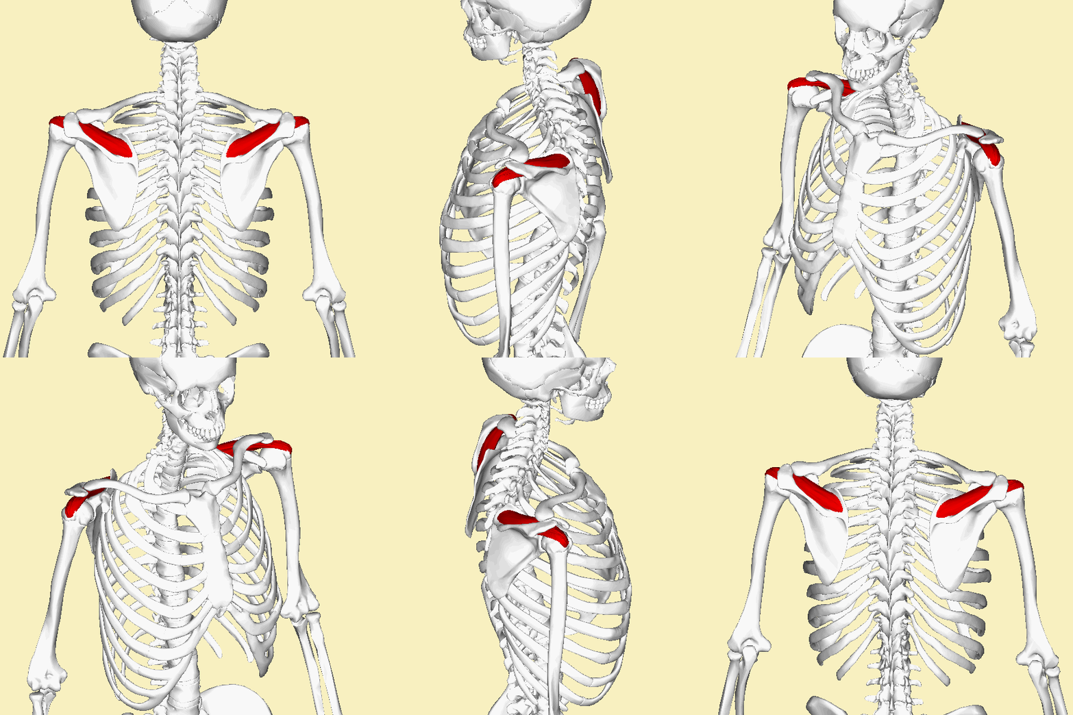
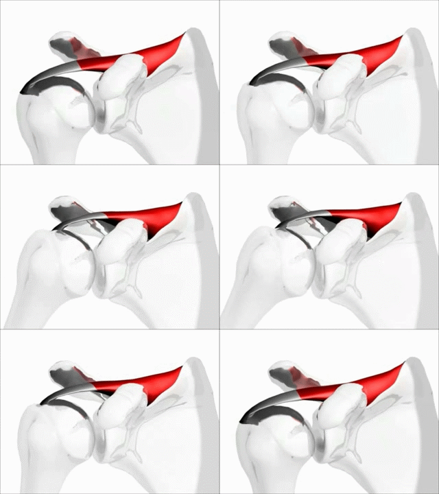
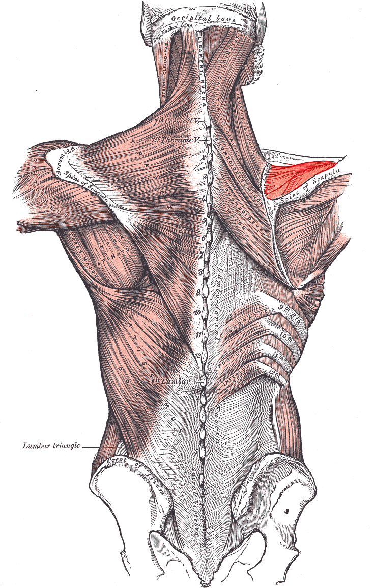
The supraspinatus (pl.: supraspinati) is a relatively small muscle of the upper back that runs from the supraspinous fossa superior portion of the scapula (shoulder blade) to the greater tubercle of the humerus. It is one of the four rotator cuff muscles and also abducts the arm at the shoulder. The spine of the scapula separates the supraspinatus muscle from the infraspinatus muscle, which originates below the spine.
Click again to return to the front
Origin
Hover to read
Click to keep this side open
Origin
Supraspinous fossa of scapula
Click again to return to the front
Insertion
Hover to read
Click to keep this side open
Insertion
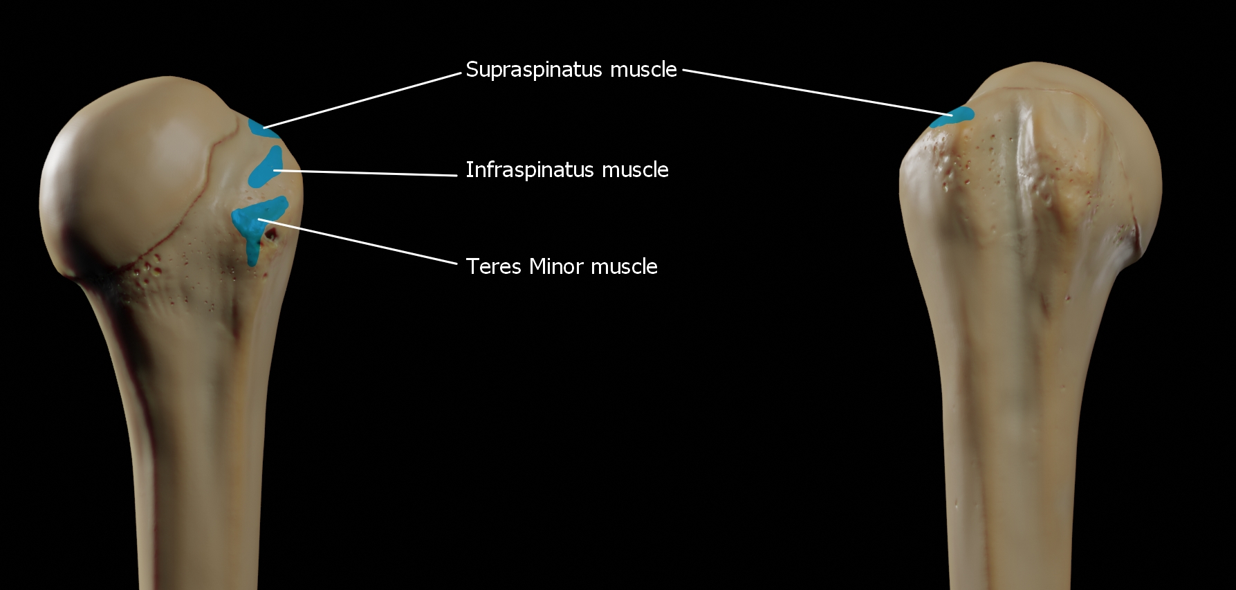
Superior facet of greater tubercle of humerus
Click again to return to the front
Actions
Hover to read
Click to keep this side open
Actions
Abduction of arm and stabilizes humerus
Click again to return to the front
Innervation
Hover to read
Click to keep this side open
Innervation
Suprascapular nerve
Click again to return to the front

Blood Supply
Hover to read
Click to keep this side open
Blood Supply
Suprascapular artery
Click again to return to the front
Palpation
Hover to read
Click to keep this side open
Palpation
Position the client comfortably and identify the muscle's bony landmarks. Ask for a gentle, pain-free contraction that matches the muscle's action, then palpate from the proximal attachment toward the distal attachment. Avoid forceful palpation over acute injury, inflamed tissue, major neurovascular structures, or areas outside the massage therapist's scope.
Click again to return to the front
Massage Applications
Hover to read
Click to keep this side open
Massage Applications
Use broad warming strokes, compression, kneading, and longitudinal work based on the muscle's size, depth, fiber direction, and client tolerance. Use focused work only after careful assessment. Avoid aggressive pressure with acute strain, suspected tear, infection, unexplained swelling, recent surgery, fracture, or other contraindications.
Click again to return to the front
Common Conditions
Hover to read
Click to keep this side open
Common Conditions
Possible concerns include strain, overuse, protective guarding, trigger-point activity, altered movement patterns, weakness, and referred discomfort. The exact clinical presentation depends on the muscle's location and function and should be evaluated within the massage therapist's scope of practice.
Click again to return to the front
Stretching
Hover to read
Click to keep this side open
Stretching
Move the related joint slowly in the direction opposite the muscle's primary action. Stabilize the proximal segment, remain within a pain-free range, and avoid bouncing or forcing the stretch.
Click again to return to the front
Strengthening
Hover to read
Click to keep this side open
Strengthening
Use low-load, controlled movement in the direction of the muscle's primary action. Begin with isometric activation when appropriate, then progress through a comfortable range while maintaining proper alignment and avoiding compensation.
Click again to return to the front
Media Credits
Attribution and licensing information for the pictures and videos used in this lesson:
- Picture 1: Supraspinatus muscle animation.gif — Anatomography — CC BY-SA 2.1 jp — Source
- Picture 2: Shoulder motion with rotator cuff (supraspinatus).gif — Young Lae, Moon M.D. Chair of 3D Based Medical Application Working group. Chairman and Professor of Orthopaedics, Chosun University Hospital, Korea. — CC BY 3.0 — Source
- Picture 3: Supraspinatus.PNG — Mikael Häggström. When using this image in external works, it may be cited as: Häggström, Mikael (2014). "Medical gallery of Mikael Häggström 2014". WikiJournal of Medicine 1 (2). DOI:10.15347/wjm/2014.008. ISSN 2002-4436. Public Domain. or By Mikael Häggström, used with permission. — Public domain — Source
- Picture 4: Attachments-of-greater-tubercle.jpg — Doctor Jana — CC BY-SA 4.0 — Source
- Picture 5: Gray — musculus supraspinatus.png — Henry Vandyke Carter — Public domain — Source