Section 01 of 20
Introduction
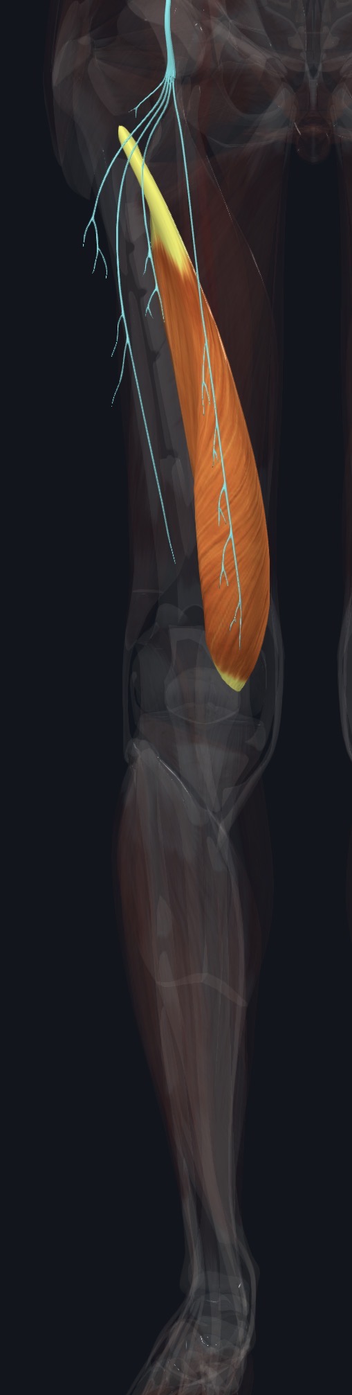
Vastus medialis lies on the inner front of the thigh and has a distinctive teardrop shape, with its lower, more oblique fibers (sometimes called vastus medialis obliquus) forming a bulge just above the inner side of the kneecap.
Along with the other three quadriceps muscles, it inserts into the quadriceps tendon around the patella. Its oblique lower fibers are especially important for pulling the kneecap medially, balancing the lateral pull of vastus lateralis and the iliotibial band.
Section 02 of 20
Origin
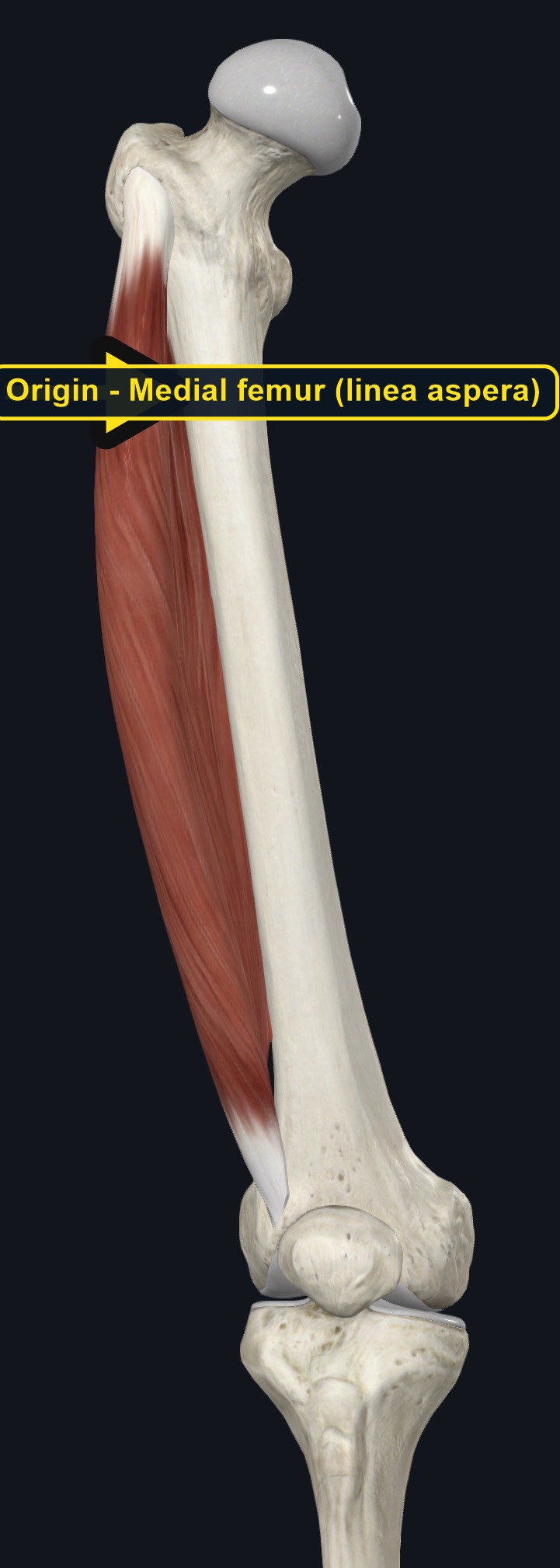
| Origin | The lower part of the intertrochanteric line, the medial lip of the linea aspera, the medial supracondylar line, and the adductor tendons of the femur. |
|---|
Section 03 of 20
Insertion
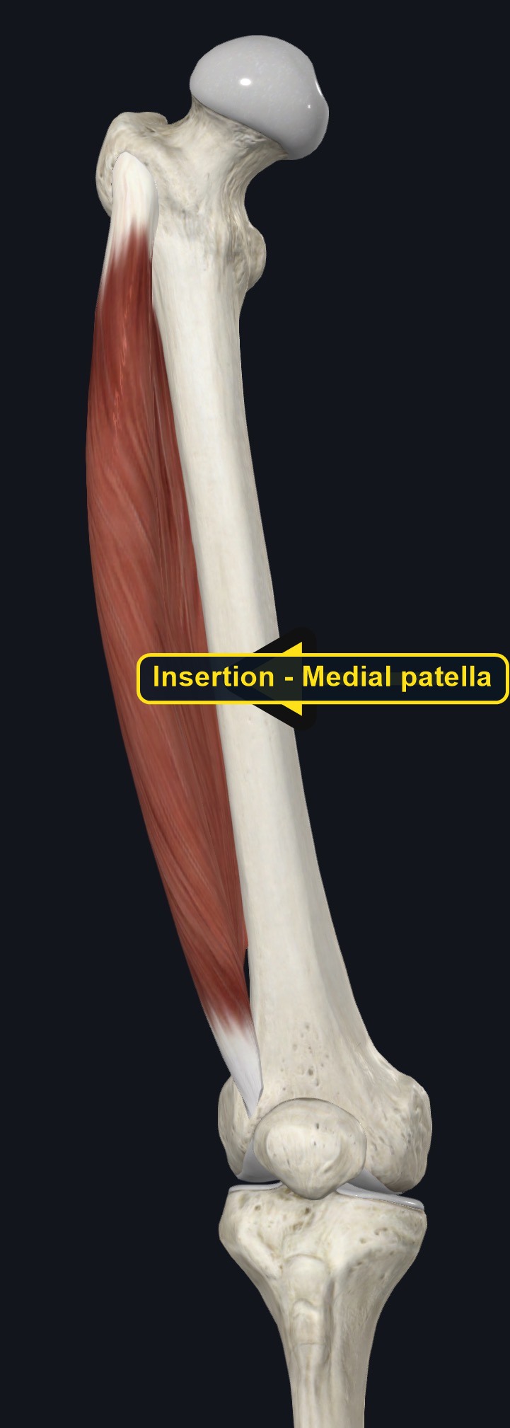
| Insertion | The medial border of the patella, and then the tibial tuberosity through the patellar ligament, joining the tendons of the other quadriceps muscles. |
|---|
Section 04 of 20
Actions
No dedicated action video is currently available. This movement is explained below and video will be added when verified source media becomes available.
| Primary action | Knee extension. |
|---|---|
| Secondary actions | Medial tracking assistance for the patella, especially in the last few degrees of extension. |
| Movement at the joint | Knee joint. |
| Heads / fiber groups | Single muscle with a distinct oblique lower portion (vastus medialis obliquus). |
| Functional examples | Standing up from sitting; walking and running; squatting; stepping down stairs with control. |
Knee extension
Straightens the knee, together with the other three quadriceps muscles.
Medial patellar tracking
The oblique lower fibers pull the kneecap medially, especially during the last few degrees of knee extension, balancing the pull of vastus lateralis.
Section 05 of 20
Nerve Innervation
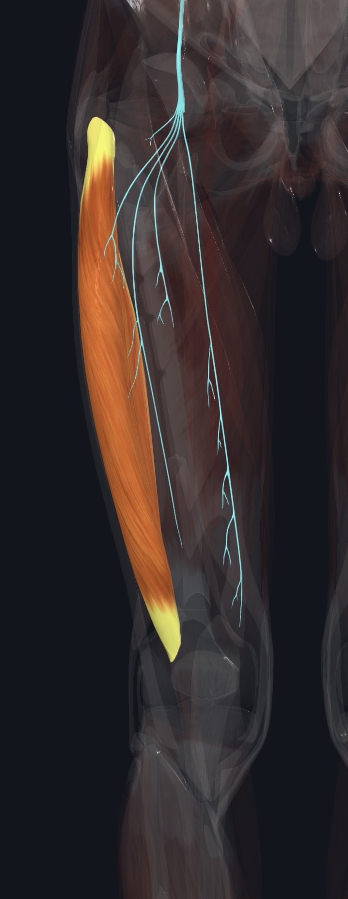
| Nerve | Femoral nerve, roots L2–L4. |
|---|
The femoral nerve supplies all four quadriceps muscles, and the saphenous nerve (a sensory branch of the femoral nerve) runs near vastus medialis toward the inner knee and leg. Numbness along the inner knee or shin, in addition to muscle weakness, may reflect nerve involvement.
Section 06 of 20
Blood Supply
| Blood | The femoral artery and the descending genicular artery, with contributions from the medial circumflex femoral artery. |
|---|
Section 07 of 20
Pictures and Visual Anatomy
Section 08 of 20
Functional Movement
Vastus medialis is active in standing up, walking, running, and especially in controlling the last part of knee straightening, such as during a squat or when stabilizing the knee while stepping down. It is often specifically targeted in rehabilitation exercises for knee and patellar problems.
Section 09 of 20
Synergists and Antagonists
Synergists: rectus femoris, vastus lateralis, and vastus intermedius (knee extension). Antagonists: the hamstrings (biceps femoris, semitendinosus, and semimembranosus), which flex the knee.
Section 10 of 20
Palpation and Location
Client position and technique
Position the client supine with the knee supported. Locate the inner side of the knee just above the patella, where the teardrop-shaped bulge of vastus medialis is often visible, then follow the muscle up the inner thigh. Ask the client to extend the knee fully, especially the last few degrees, to feel the oblique lower fibers engage.
Protect nearby structures
Use moderate pressure over the muscle belly, and lighter pressure near the knee. Avoid deep pressure over the femoral triangle at the top of the thigh, and stop if the client reports sharp pain, numbness, or tingling.
Section 11 of 20
Massage Therapy Relevance
Vastus medialis, especially its oblique fibers, is central to healthy kneecap tracking, and its weakness is commonly discussed in patellofemoral pain and after knee injury or surgery. It is one of the first quadriceps muscles to show visible wasting after knee problems.
Section 12 of 20
Dysfunction: What Happens When Movement Goes Wrong
Changes in how a muscle works can change how a joint moves. The points below describe possibilities that may be associated with altered muscle function; they are not diagnoses, and one muscle is never assumed to be the sole cause of a problem.
When the muscle is weak
Weakness, especially of the oblique fibers, may be associated with poor kneecap tracking and patellofemoral pain.
When it is tight or shortened
Tightness is less commonly discussed than weakness for this muscle, but may be associated with general anterior and medial thigh tightness.
When it is overused
Rehabilitation exercises or activities focused on the last degrees of knee extension may temporarily fatigue the muscle.
When it is strained
A strain may cause pain on the inner thigh with resisted knee extension; knee joint and ligament problems can look similar.
When it is inhibited or others compensate
Reduced or delayed activation relative to vastus lateralis is commonly discussed in patellofemoral pain and after knee injury or effusion.
When it does not coordinate well with synergists and antagonists
Poor coordination or timing relative to vastus lateralis may alter kneecap tracking and may be associated with patellofemoral symptoms.
Section 13 of 20
Conditions, Pathologies, and Pain Relationships
Patellofemoral pain syndrome
Weakness or delayed activation of vastus medialis relative to vastus lateralis may be associated with poor kneecap tracking and pain around or under the kneecap.
Quadriceps weakness after knee injury or surgery
Vastus medialis is often one of the first muscles to visibly weaken and atrophy after knee injury, effusion, or surgery.
Quadriceps strain
Sudden forceful contraction can strain the muscle, causing pain on the inner thigh with resisted knee extension.
Differential considerations
Knee joint problems, meniscus injury, medial collateral ligament injury, and referred pain from the hip can all cause similar inner thigh and knee symptoms.
Section 14 of 20
Example Clinical Relationship
A runner reports a nagging ache around the kneecap that worsens with downhill running and prolonged sitting. Vastus medialis weakness relative to vastus lateralis may be involved, and the pattern may be associated with patellofemoral pain syndrome, but meniscus and ligament injuries can look similar. Gentle work on the quadriceps as a group may support comfort, while a knee that gives way and swells, or sudden severe swelling after injury, needs referral.
Section 15 of 20
Massage and Treatment Approaches
For healthy, non-acute tissue, position the client supine with the knee supported. Begin with broad effleurage from the knee toward the hip, then use kneading and compression along the muscle, and gentle work over the teardrop-shaped area above the kneecap within comfortable pressure.
| Movement | Use gentle active knee extension with attention to the last few degrees of straightening, within comfort. |
|---|---|
| Regional work | Consider rectus femoris, vastus lateralis, the adductors, and the inner knee structures when assessment supports it. |
| Avoid | Deep pressure over the femoral triangle, over an acute strain, or at any point that reproduces leg tingling. |
Section 16 of 20
Precautions and Contraindications
Precautions and contraindications
- Acute strain, recent hip, thigh, or knee injury or surgery.
- Use caution with anticoagulant use, easy bruising, or varicose veins.
- Avoid deep pressure over the femoral triangle.
- Stop if pressure causes sharp pain, numbness, or tingling.
Section 17 of 20
Red Flags and When to Refer
Red flags and referral
- A knee that repeatedly gives way, locks, or swells significantly after activity.
- A swollen, warm, red, or painful leg, especially on one side (possible blood clot).
- Sudden severe thigh or knee pain and swelling after an injury, with inability to bear weight.
- Thigh or knee pain with fever, night pain, or unexplained weight loss.
Section 18 of 20
Study Summary
Attachments
Origin: intertrochanteric line, linea aspera, and medial supracondylar line of the femur. Insertion: medial patella, then the tibial tuberosity.
Actions
Extends the knee and helps guide the kneecap medially.
Supply
Femoral nerve (L2–L4); femoral and descending genicular arteries.
Section 19 of 20
Instructor Narration
Open narration transcript
Vastus medialis is the teardrop-shaped quadriceps muscle on the inner front of the thigh, with a bulge just above the inner kneecap.
It arises from the intertrochanteric line, the linea aspera, and the medial supracondylar line of the femur, and inserts on the medial border of the patella, then continues to the tibial tuberosity.
It extends the knee and its oblique lower fibers pull the kneecap medially, balancing vastus lateralis. Rectus femoris and vastus intermedius help it, and the hamstrings oppose it.
The femoral nerve supplies it, from L2 to L4, and the femoral and descending genicular arteries provide its blood.
Palpate the teardrop-shaped bulge just above the inner knee and follow it up the inner thigh, asking the client to fully straighten the knee to feel the oblique fibers engage. Avoid deep pressure over the femoral triangle.
Patellofemoral pain, post-injury quadriceps weakness, and strain are common, and knee joint, meniscus, and ligament problems can overlap. Refer a knee that repeatedly gives way or swells, a swollen warm leg, sudden severe swelling after injury, or thigh or knee pain with fever.
Remember: intertrochanteric line and linea aspera to the medial patella and tibial tuberosity; knee extension and medial patellar tracking; femoral nerve.
Section 20 of 20