Section 01 of 20
Introduction
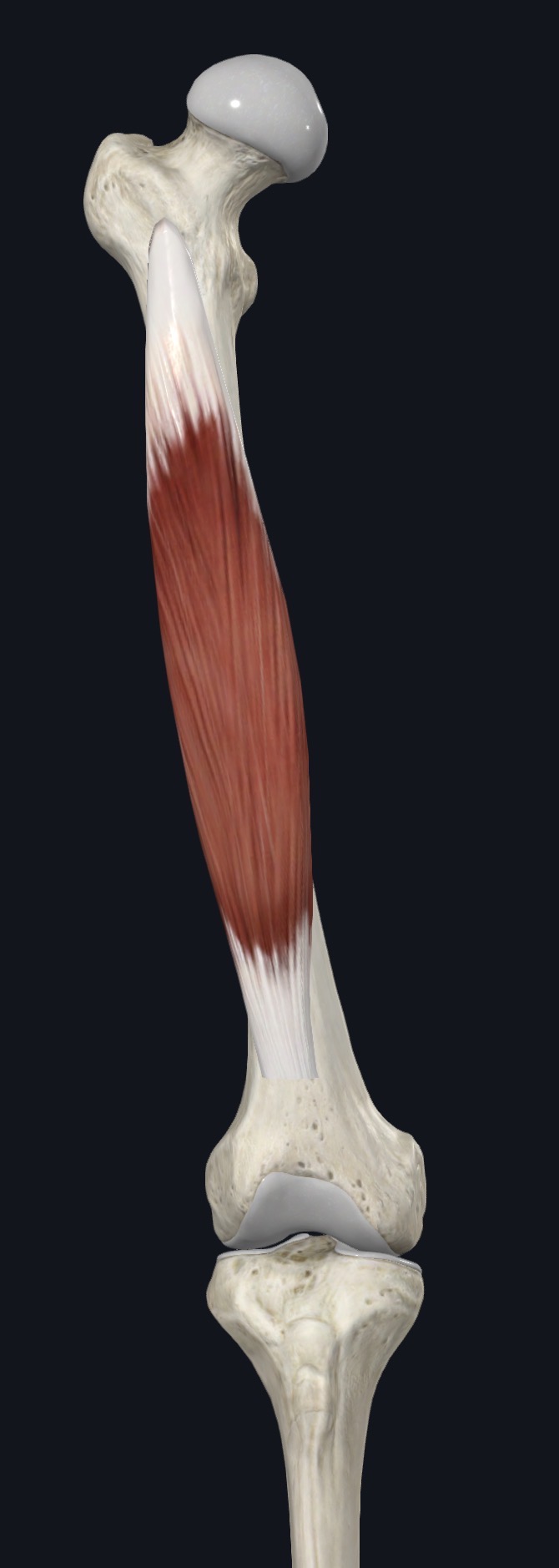
Vastus intermedius lies directly on the front and side of the femur, deep to rectus femoris, in the middle of the anterior thigh. It cannot be seen or felt separately from the surface because rectus femoris covers almost all of it.
It is one of the four muscles of the quadriceps group, along with rectus femoris, vastus lateralis, and vastus medialis. Unlike rectus femoris, it crosses only the knee joint, not the hip, so its only job is to straighten the knee.
Section 02 of 20
Origin
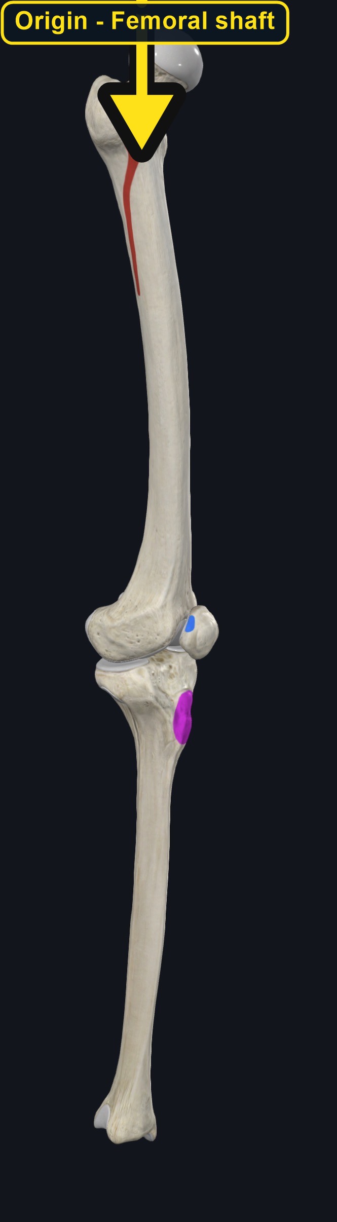
| Origin | The anterior and lateral surfaces of the upper two-thirds of the shaft of the femur. |
|---|
Section 03 of 20
Insertion
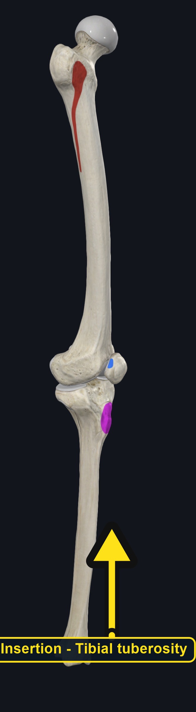
| Insertion | The base of the patella, and then the tibial tuberosity through the patellar ligament, joining the tendons of the other quadriceps muscles. |
|---|
Section 04 of 20
Actions
No dedicated action video is currently available. This movement is explained below and video will be added when verified source media becomes available.
| Primary action | Knee extension. |
|---|---|
| Secondary actions | None; it has no action at the hip. |
| Movement at the joint | Knee joint only. |
| Heads / fiber groups | Single muscle, sometimes closely blended with vastus lateralis. |
| Functional examples | Standing up from sitting; walking and running; climbing stairs; kicking. |
Knee extension
Straightens the knee, together with the other three quadriceps muscles.
Section 05 of 20
Nerve Innervation
| Nerve | Femoral nerve, roots L2–L4. |
|---|
The femoral nerve supplies all four quadriceps muscles. Weakness of knee extension with a knee that buckles, especially after a thigh or hip injury, may reflect femoral nerve involvement, not only muscle weakness.
Section 06 of 20
Blood Supply
| Blood | Branches of the femoral artery and the lateral circumflex femoral artery. |
|---|
Section 07 of 20
Pictures and Visual Anatomy
Section 08 of 20
Functional Movement
Vastus intermedius works with the rest of the quadriceps in every activity that straightens the knee: standing up, walking, running, climbing stairs, and kicking. Because it has no role at the hip, it contributes purely to knee extension strength.
Section 09 of 20
Synergists and Antagonists
Synergists: rectus femoris, vastus lateralis, and vastus medialis (knee extension). Antagonists: the hamstrings (biceps femoris, semitendinosus, and semimembranosus), which flex the knee.
Section 10 of 20
Palpation and Location
Client position and technique
Vastus intermedius cannot be felt directly because it lies completely beneath rectus femoris. Position the client supine with the knee supported. Gently move rectus femoris aside or work through it with broad, moderate pressure along the center of the front of the thigh, and ask for a straight leg raise or knee extension to appreciate the deeper layer contracting beneath the surface muscle.
Protect nearby structures
Because direct palpation is not possible, avoid excessive pressure trying to reach this muscle. Stay below the inguinal crease and off the femoral triangle at the top of the thigh, and stop if the client reports sharp pain, numbness, or tingling.
Section 11 of 20
Massage Therapy Relevance
Vastus intermedius contributes to overall quadriceps strength and is relevant in knee rehabilitation and after knee surgery, when quadriceps weakness (particularly of the vasti) is common. Because it cannot be felt separately, it is typically addressed through general quadriceps work.
Section 12 of 20
Dysfunction: What Happens When Movement Goes Wrong
Changes in how a muscle works can change how a joint moves. The points below describe possibilities that may be associated with altered muscle function; they are not diagnoses, and one muscle is never assumed to be the sole cause of a problem.
When the muscle is weak
Weakness may reduce knee extension strength and is common after knee injury or surgery, contributing to a sense of the knee giving way.
When it is tight or shortened
Tightness is difficult to isolate from rectus femoris and may be associated with general anterior thigh tightness and limited knee flexion.
When it is overused
Repeated knee extension in running, jumping, and cycling may overload the quadriceps group, including vastus intermedius.
When it is strained
A strain is uncommon in isolation and usually occurs as part of a broader quadriceps strain; knee joint problems can look similar.
When it is inhibited or others compensate
Reduced activation, along with vastus medialis, is common after knee injury or effusion and may be associated with quadriceps weakness.
When it does not coordinate well with synergists and antagonists
Poor coordination with the other quadriceps muscles may alter knee control and may be associated with patellofemoral or knee symptoms.
Section 13 of 20
Conditions, Pathologies, and Pain Relationships
Quadriceps weakness after knee injury or surgery
Vastus intermedius, along with vastus medialis, is often inhibited after knee injury or surgery (such as after ACL reconstruction), contributing to reduced knee extension strength.
Quadriceps contusion
A direct blow to the front of the thigh can bruise the deep muscle layers, including vastus intermedius, and severe bruising needs medical assessment.
Quadriceps tendinopathy
Overuse of the shared quadriceps tendon may cause pain above the kneecap; this involves the whole quadriceps group, not vastus intermedius alone.
Differential considerations
Knee joint problems, patellofemoral pain, femoral nerve entrapment, and hip joint referral can all cause similar anterior thigh and knee symptoms.
Section 14 of 20
Example Clinical Relationship
A client recovering from knee surgery reports ongoing thigh weakness and a knee that feels unstable when going down stairs, even though the muscle appears less visibly wasted than before. Vastus intermedius and vastus medialis are often inhibited after knee surgery, and the pattern may be associated with post-surgical quadriceps weakness, but ongoing joint or nerve problems can look similar. Gentle work on the quadriceps as a group may support comfort, while a hard lump, a swollen warm leg, or worsening instability needs referral.
Section 15 of 20
Massage and Treatment Approaches
For healthy, non-acute tissue, position the client supine with the knee supported. Begin with broad effleurage along the front of the thigh, then use kneading and compression through rectus femoris to reach the deeper layer, working within comfortable pressure.
| Movement | Use gentle passive and active knee extension and flexion within comfort; do not force range. |
|---|---|
| Regional work | Consider rectus femoris, vastus lateralis, vastus medialis, and the iliotibial band when assessment supports it. |
| Avoid | Deep pressure in the femoral triangle, over an acute contusion, or at any point that reproduces leg tingling. |
Section 16 of 20
Precautions and Contraindications
Precautions and contraindications
- Acute strain, a recent deep thigh bruise, or recent hip, thigh, or knee injury or surgery.
- Use caution with anticoagulant use, easy bruising, or a history of thigh trauma with a hard lump.
- Avoid deep pressure over the femoral triangle.
- Stop if pressure causes sharp pain, numbness, or tingling.
Section 17 of 20
Red Flags and When to Refer
Red flags and referral
- A hard, painful lump in the thigh after an injury that is not settling (possible myositis ossificans).
- A swollen, warm, red, or painful leg, especially on one side (possible blood clot).
- Sudden severe thigh pain and swelling after a direct blow, with inability to bear weight.
- Thigh pain with fever, night pain, or unexplained weight loss.
Section 18 of 20
Study Summary
Attachments
Origin: anterior and lateral shaft of the femur. Insertion: patella, then the tibial tuberosity.
Actions
Extends the knee.
Supply
Femoral nerve (L2–L4); femoral and lateral circumflex femoral arteries.
Section 19 of 20
Instructor Narration
Open narration transcript
Vastus intermedius is the deepest quadriceps muscle, hidden completely beneath rectus femoris on the front of the thigh.
It arises from the anterior and lateral surfaces of the femoral shaft and inserts on the patella, then continues to the tibial tuberosity through the patellar ligament.
Its only action is knee extension, working together with the other three quadriceps muscles. The hamstrings oppose it.
The femoral nerve supplies it, from L2 to L4, and branches of the femoral and lateral circumflex femoral arteries provide its blood.
It cannot be felt separately from rectus femoris. Work through the surface muscle with broad, moderate pressure along the center of the thigh, and ask for knee extension to appreciate the deeper contraction.
Quadriceps weakness after knee injury or surgery, contusion, and tendinopathy are relevant, and knee joint and nerve problems can overlap. Refer a hard lump after injury that is not settling, a swollen warm leg, sudden severe thigh swelling with inability to bear weight, or thigh pain with fever.
Remember: femoral shaft to the patella and tibial tuberosity; knee extension only; femoral nerve.
Section 20 of 20