Section 01 of 20
Introduction
Tibialis anterior is a long, spindle-shaped muscle that lies just to the outer side of the shin bone (tibia) and is the most superficial muscle of the anterior leg compartment. Its firm belly can be felt easily along the front of the lower leg.
Its long tendon crosses the front of the ankle and travels to the inner side of the foot, which is why the muscle both lifts the foot and turns the sole slightly inward.
Section 02 of 20
Origin
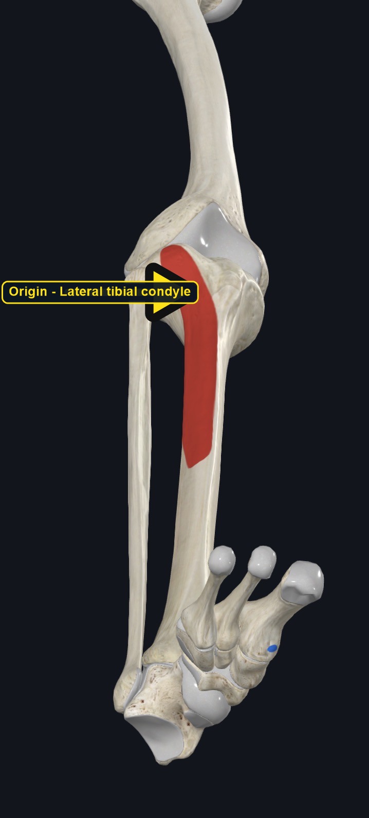
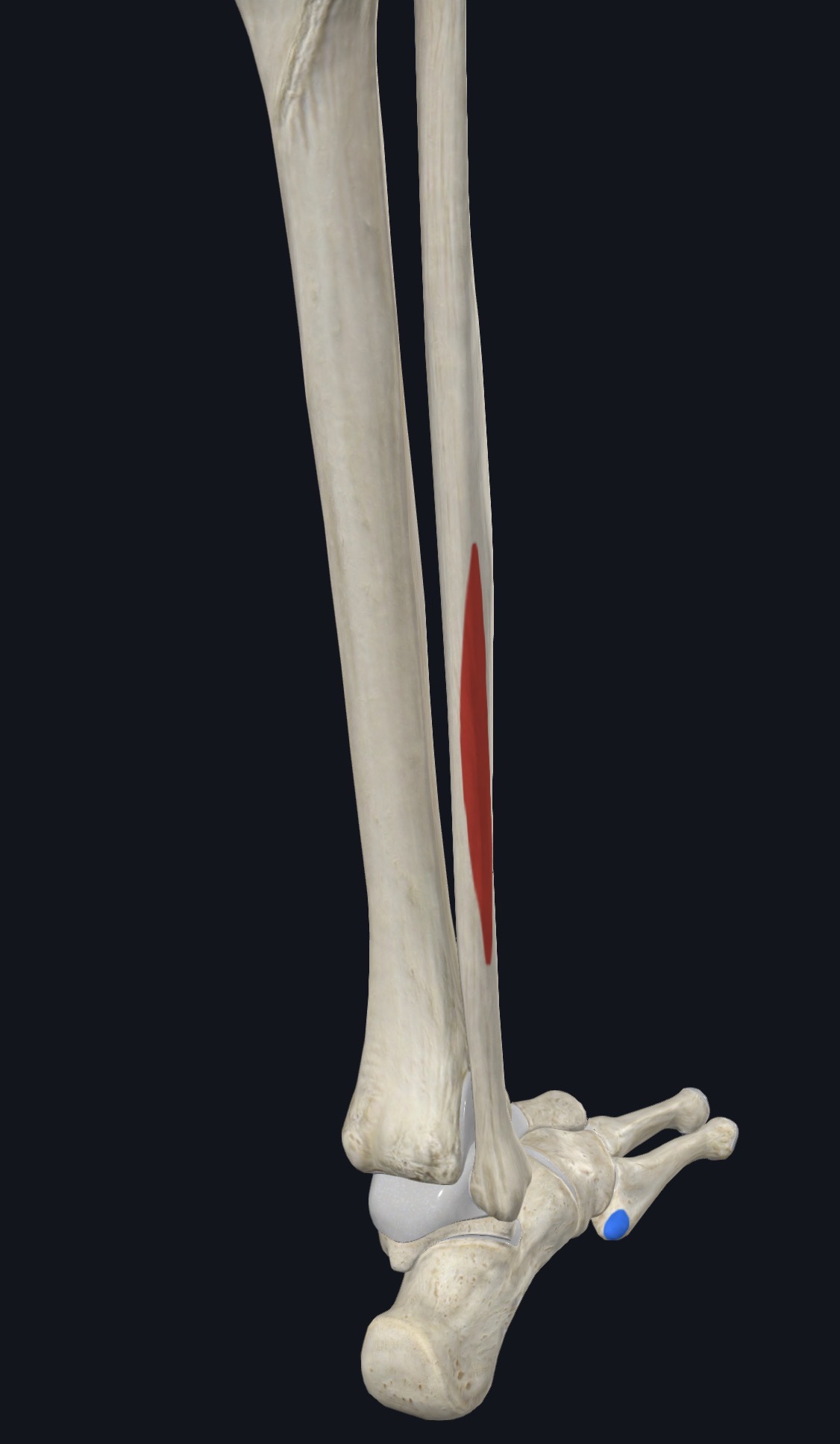
| Origin | The lateral condyle of the tibia and the upper half to two-thirds of the lateral surface of the tibial shaft, along with the adjacent interosseous membrane. |
|---|
Section 03 of 20
Insertion
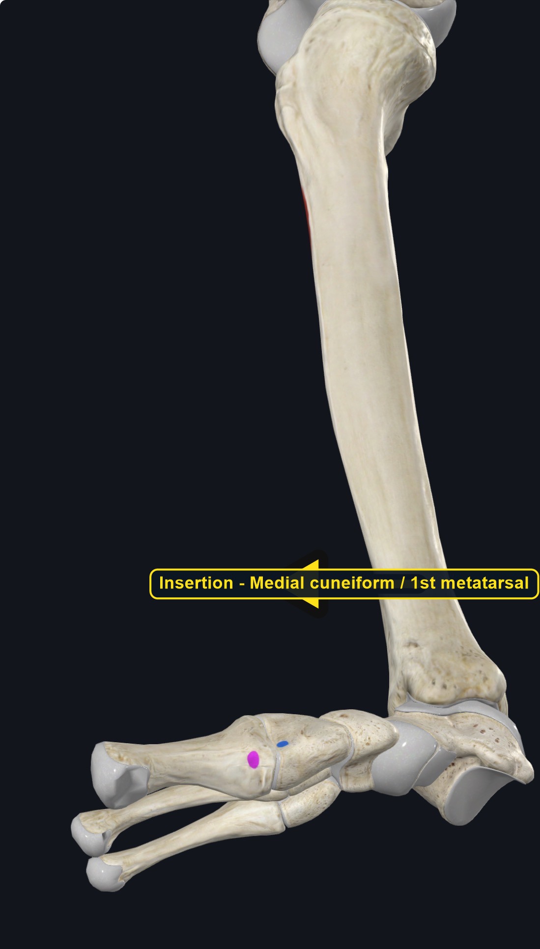
| Insertion | The medial cuneiform bone and the base of the first metatarsal, on the plantar (sole) side. |
|---|
Section 04 of 20
Actions
| Primary action | Dorsiflexion of the ankle. |
|---|---|
| Secondary actions | Inversion of the foot and control of foot clearance during gait. |
| Movement at the joint | Ankle (talocrural) and subtalar joints. |
| Heads / fiber groups | Single spindle-shaped muscle. |
| Functional examples | Walking and running (swing phase toe clearance and controlled heel-strike lowering); walking on the heels; pulling the toes up. |
Dorsiflexion of the ankle
Lifts the foot up toward the shin.
Inversion of the foot
Turns the sole of the foot inward.
Foot clearance during walking
Controls the lowering of the foot after heel strike and lifts the toes during the swing phase of walking, preventing the foot from dragging.
Section 05 of 20
Nerve Innervation
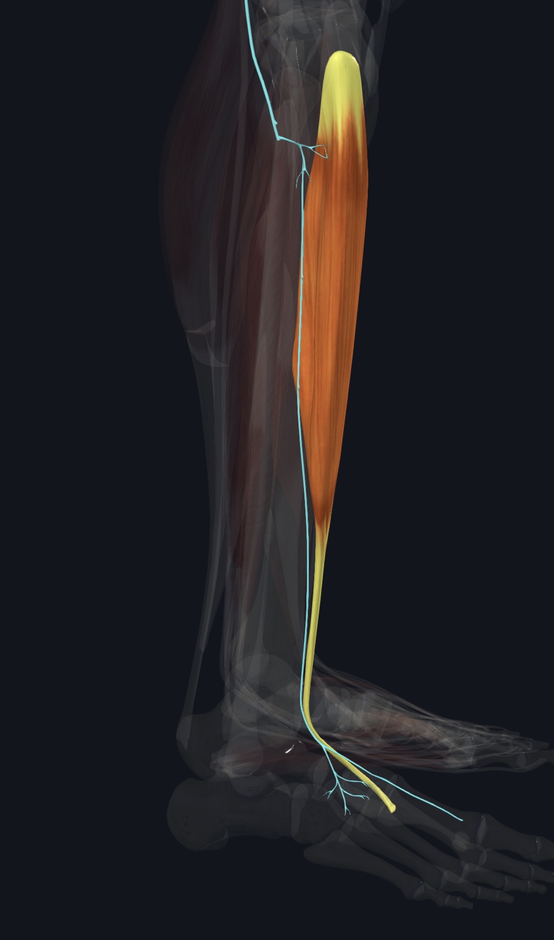
| Nerve | Deep fibular (peroneal) nerve, roots L4–S1. |
|---|
The deep fibular nerve and anterior tibial vessels run along the deep side of the muscle. Weakness of dorsiflexion causing a foot drop, or numbness in the web space between the first and second toes, may reflect nerve involvement, not only muscle tightness.
Section 06 of 20
Blood Supply
| Blood | The anterior tibial artery. |
|---|
Section 07 of 20
Pictures and Visual Anatomy
Section 08 of 20
Functional Movement
Tibialis anterior is essential for a normal walking and running gait, lifting the toes during the swing phase so the foot clears the ground, and controlling the foot as the heel touches down. It is also active in activities like walking on the heels or pulling the toes upward.
Section 09 of 20
Synergists and Antagonists
Synergists: extensor hallucis longus and extensor digitorum longus (dorsiflexion), and tibialis posterior (inversion). Antagonists: gastrocnemius and soleus (plantar flexion), and fibularis longus and brevis (eversion).
Section 10 of 20
Palpation and Location
Client position and technique
Position the client supine with the knee slightly bent and the foot relaxed. Locate the bony ridge of the tibia and feel just lateral to it for the firm muscle belly. Ask the client to dorsiflex and invert the foot to feel the muscle and its tendon stand out clearly along the front of the ankle.
Protect nearby structures
Use moderate pressure over the muscle belly and lighter pressure over the tendon at the ankle and instep, where it lies close to bone with little padding. Avoid deep pressure directly over the anterior tibial vessels and nerve, and stop if the client reports sharp pain, numbness, or tingling.
Section 11 of 20
Massage Therapy Relevance
Tibialis anterior is commonly involved in shin pain from running and walking on hard surfaces, and its tightness or weakness can affect gait. Shin pain has several possible causes, so careful assessment is useful.
Section 12 of 20
Dysfunction: What Happens When Movement Goes Wrong
Changes in how a muscle works can change how a joint moves. The points below describe possibilities that may be associated with altered muscle function; they are not diagnoses, and one muscle is never assumed to be the sole cause of a problem.
When the muscle is weak
Weakness may cause a foot drop or slapping gait, with the foot not clearing the ground properly during walking.
When it is tight or shortened
Tightness may be associated with limited plantar flexion and a feeling of tightness along the front of the shin.
When it is overused
Running, walking on hard or uneven surfaces, and hiking downhill may overload the muscle and may contribute to shin splints.
When it is strained
A strain may cause pain along the front of the shin with resisted dorsiflexion; stress fracture and compartment syndrome must be ruled out.
When it is inhibited or others compensate
Reduced activation may be associated with a slapping gait and more reliance on hip flexion to clear the foot.
When it does not coordinate well with synergists and antagonists
Poor coordination with the calf muscles and the other dorsiflexors may alter gait mechanics and may be associated with shin or ankle symptoms.
Section 13 of 20
Conditions, Pathologies, and Pain Relationships
Anterior shin splints
Repetitive impact, especially downhill walking or running, may overload the muscle at its attachment to the tibia and cause pain along the front of the shin.
Tibialis anterior tendinopathy
Overuse may irritate the tendon at the front of the ankle, causing pain with walking or dorsiflexion.
Anterior compartment syndrome
Swelling within the tight anterior compartment, sometimes after intense exercise or injury, can compress the muscle, nerve, and vessels and is a medical emergency if acute.
Differential considerations
Stress fractures of the tibia, deep fibular nerve entrapment, and referred pain from the lumbar spine can all cause similar shin and foot symptoms.
Section 14 of 20
Example Clinical Relationship
A new runner reports pain along the front of the shin that builds over a run and eases with rest. Tibialis anterior may be involved, and the pattern may be associated with shin splints from a sudden increase in training, but a stress fracture or early compartment syndrome can look similar. Gentle work along the muscle belly may support comfort, while severe worsening pain and tightness after exercise needs urgent referral.
Section 15 of 20
Massage and Treatment Approaches
For healthy, non-acute tissue, position the client supine with the knee slightly bent and supported. Begin with light effleurage along the front of the shin, then use gentle kneading of the muscle belly and lighter strokes over the tendon at the ankle, staying within comfortable pressure.
| Movement | Use gentle ankle plantar flexion and eversion stretches within comfort; do not force range. |
|---|---|
| Regional work | Consider extensor digitorum longus, extensor hallucis longus, and the calf muscles when assessment supports it. |
| Avoid | Deep pressure directly over the shin bone or the anterior tibial vessels, and any point that reproduces sharp pain, numbness, or tingling. |
Section 16 of 20
Precautions and Contraindications
Precautions and contraindications
- Acute shin injury, suspected stress fracture, or recent leg or ankle surgery.
- Never work over a leg with symptoms of compartment syndrome: severe swelling, tightness, and pain out of proportion to injury.
- Use caution with anticoagulant use or easy bruising.
- Stop if pressure causes sharp pain, numbness, or tingling.
Section 17 of 20
Red Flags and When to Refer
Red flags and referral
- Severe, worsening leg pain and tightness after exercise or injury, especially with numbness or pain on passive toe stretch (possible compartment syndrome; this is an emergency).
- A foot drop, or new difficulty lifting the foot or toes.
- Point tenderness on the shin bone that worsens with activity and does not improve with rest (possible stress fracture).
- A swollen, warm, red, or painful leg on one side (possible blood clot).
Section 18 of 20
Study Summary
Attachments
Origin: lateral tibial condyle and shaft. Insertion: medial cuneiform and first metatarsal base.
Actions
Dorsiflexion and inversion of the foot.
Supply
Deep fibular nerve (L4–S1); anterior tibial artery.
Section 19 of 20
Instructor Narration
Open narration transcript
Tibialis anterior is the prominent muscle along the front of the shin, just lateral to the tibia.
It arises from the lateral tibial condyle and the upper lateral shaft of the tibia, and inserts on the medial cuneiform and the base of the first metatarsal.
It dorsiflexes and inverts the foot, and controls foot clearance during walking. Extensor digitorum longus and extensor hallucis longus help it, and gastrocnemius and soleus oppose it.
The deep fibular nerve supplies it, from L4 to S1, and the anterior tibial artery provides its blood.
Palpate lateral to the tibial ridge, and ask for dorsiflexion and inversion to feel the muscle and its tendon stand out at the ankle. Use lighter pressure over the tendon.
Shin splints, tendinopathy, and stress fractures are common, and compartment syndrome is a medical emergency to screen for. Refer severe worsening leg pain and tightness after exercise, a new foot drop, point tenderness that worsens with activity, or a swollen warm leg.
Remember: lateral tibial condyle and shaft to the medial cuneiform and first metatarsal; dorsiflexion and inversion; deep fibular nerve.
Section 20 of 20