Section 01 of 20
Introduction
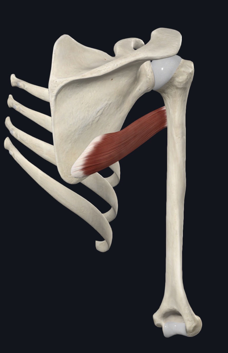
Teres major is a thick, somewhat cylindrical ("teres" means round/rope-like in Latin) muscle running along the lower/lateral border of the scapula to the humerus. Despite lying immediately adjacent to the rotator cuff muscles, teres major is not itself part of the rotator cuff — it is a distinct, more superficial muscle with different innervation and a different functional role.
Teres major is often introduced to students alongside latissimus dorsi because the two muscles share very similar actions and a closely related insertion area on the humerus, even though they arise from different origins and are innervated by different nerves. This shared action pattern gives teres major its informal nickname, "lat's little helper."
For massage therapists, teres major is relevant both as a commonly tight muscle in clients who do a lot of pulling, climbing, or swimming-type activity, and as an important structure to distinguish from its neighbor, teres minor, which is a true rotator cuff muscle with the opposite rotational action.
Section 02 of 20
Origin
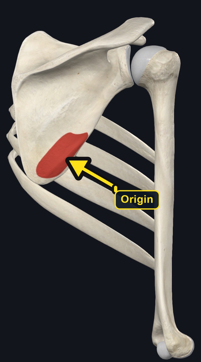
| Origin | Posterior surface of the inferior angle of the scapula (and the adjacent lower lateral border). |
|---|
Section 03 of 20
Insertion
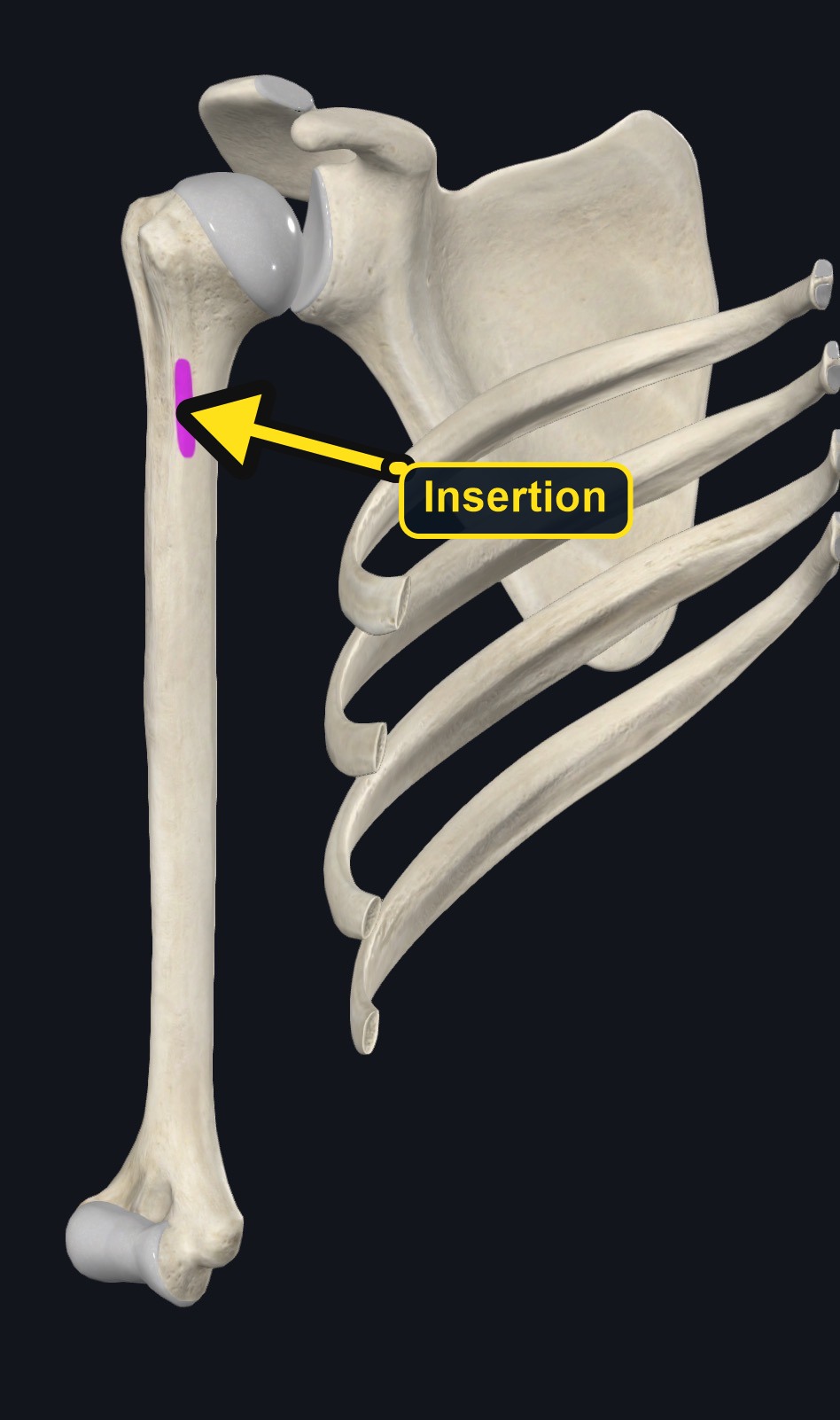
| Insertion | Medial lip of the intertubercular (bicipital) groove of the humerus — immediately adjacent to, but on the opposite lip from, the insertion of latissimus dorsi, which attaches to the floor of the same groove. |
|---|
Section 04 of 20
Actions
No dedicated action video is currently available. This movement is explained below and video will be added when verified source media becomes available.
| Primary action | Shoulder internal rotation and adduction. |
|---|---|
| Secondary actions | Extension of the arm from a flexed position; stabilizes the inferior angle of the scapula. Teres major is not a rotator cuff muscle. |
| Movement at the joint | Glenohumeral (shoulder) joint. |
| Heads / fiber groups | Single muscle belly; no separate heads. |
| Functional examples | Pulling down; rowing; climbing; swimming; placing the hand behind the back. |
Primary actions
- Internal (medial) rotation of the shoulder.
- Adduction of the shoulder.
- Extension of the shoulder (from a flexed position).
Functional note
These three actions closely mirror those of latissimus dorsi, which is why the two muscles are frequently discussed together and why teres major is sometimes informally called latissimus dorsi's "little helper," despite the two muscles having entirely separate origins and nerve supplies.
Section 05 of 20
Nerve Innervation
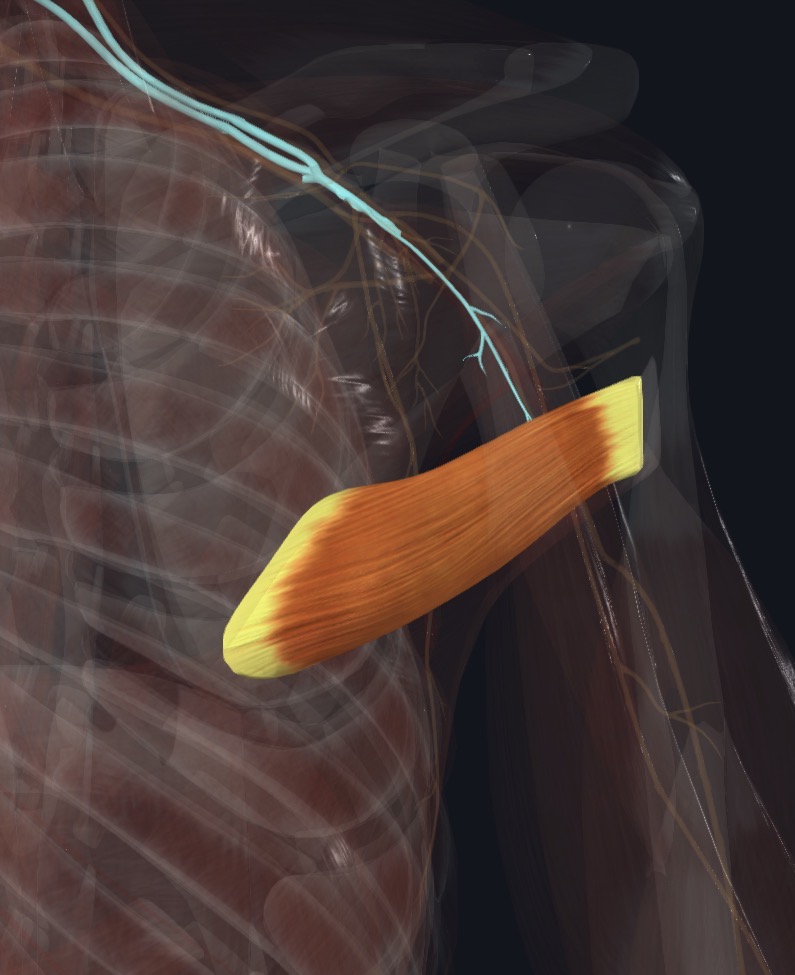
| Nerve | Lower subscapular nerve is the standard primary supply; thoracodorsal contribution is described in some references |
|---|---|
| Roots | Most commonly C5-C6; C7 contribution is variably reported |
| Relationship | The lower subscapular nerve arises from the posterior cord of the brachial plexus and supplies both teres major and the lower portion of subscapularis, which is a useful anatomical link to remember between these two muscles. |
Section 06 of 20
Blood Supply
Teres major is primarily supplied by the subscapular artery and its branch, the thoracodorsal artery, along with contribution from the circumflex scapular artery.
Section 07 of 20
Pictures and Visual Anatomy
Section 08 of 20
Functional Movement
Teres major helps pull the arm down and back and turn it inward. It works with latissimus dorsi in climbing, rowing, swimming, and pulling down, and it stabilizes the shoulder blade while the arm works.
Section 09 of 20
Synergists and Antagonists
Synergists
Latissimus dorsi (internal rotation, adduction, extension); posterior deltoid and long head of triceps assist extension.
Antagonists
Infraspinatus and teres minor (external rotation, the opposite rotational action); deltoid (abduction, opposing adduction); anterior deltoid and pectoralis major (flexion, opposing extension).
Section 10 of 20
Palpation and Location
Client position
Client prone or side-lying, arm relaxed or gently supported.
Landmarks and technique
- Locate the inferior angle of the scapula; teres major runs from this area diagonally up and out toward the humerus, forming part of the posterior border of the axilla along with latissimus dorsi.
- Teres major and latissimus dorsi are closely related in this region and can be difficult to distinguish precisely by palpation alone; asking the client to gently perform shoulder extension and internal rotation (e.g., pressing the arm down and back against light resistance) will help the therapist feel the general muscle group contract.
- Teres minor lies just superior to teres major along the lateral scapular border, and the two can sometimes be distinguished by testing the opposite action: teres minor contracts with resisted external rotation, while teres major contracts with resisted internal rotation.
- This region is close to the axilla and its neurovascular contents; palpation should remain gentle and gradual, particularly as the therapist's hand moves toward the armpit.
Section 11 of 20
Massage Therapy Relevance
Teres major is commonly involved in general tightness related to pulling, climbing, swimming, and overhead activities that combine shoulder extension and internal rotation — the same movement pattern used heavily by latissimus dorsi. Clients who do significant upper-body pulling exercise (such as rowing or pull-ups) or repetitive swimming strokes may develop noticeable tightness through this region.
Because teres major sits at the posterior border of the axilla, alongside latissimus dorsi, general tension here can also be discussed in relation to overall shoulder mobility, particularly restricted shoulder flexion or external rotation range if the muscle is significantly shortened.
Distinguishing teres major tightness from true rotator cuff (teres minor, infraspinatus, supraspinatus, subscapularis) involvement is a useful clinical skill, since the treatment considerations and injury implications can differ between a straightforward muscle tightness pattern and true rotator cuff pathology.
Section 12 of 20
Dysfunction: What Happens When Movement Goes Wrong
Changes in how a muscle works can change how a joint moves. The points below describe possibilities that may be associated with altered muscle function; they are not diagnoses, and one muscle is never assumed to be the sole cause of a problem.
When the muscle is weak
Weakness may reduce pulling and adduction strength and may shift work to latissimus dorsi and the posterior shoulder muscles.
When it is tight or shortened
Tightness may be associated with limited arm elevation and external rotation and may contribute to tension along the lower border of the scapula and the back of the armpit.
When it is overused
Repetitive pulling, rowing, and swimming may overload the muscle and may contribute to a posterior shoulder ache.
When it is strained
A strain may cause pain at the back of the armpit with resisted adduction or internal rotation; nerve and joint problems can look similar.
When it is inhibited or others compensate
Reduced activation may be associated with more reliance on latissimus dorsi and altered scapular control.
When it does not coordinate well with synergists and antagonists
Poor coordination with latissimus dorsi, the rotator cuff, and the rhomboids may alter shoulder mechanics and scapular position.
Section 13 of 20
Conditions, Pathologies, and Pain Relationships
Teres major strain / overuse tension
What it is: Muscle strain or general fatigue-related tightness, typically from repetitive or forceful pulling, climbing, or throwing-type activity.
Why the muscle may be involved: High demand during combined internal rotation, adduction, and extension movements.
Typical symptoms: Posterior shoulder/axillary border discomfort, sometimes aggravated by overhead reaching or resisted pulling movements.
Similar-presenting conditions: Latissimus dorsi strain, teres minor or infraspinatus-related symptoms, general posterior shoulder tightness.
Refer when: Pain is severe, persistent, or associated with significant weakness.
"Quadrilateral space syndrome" (informational, uncommon)
What it is: A relatively uncommon condition involving compression of the axillary nerve and posterior circumflex humeral artery within the quadrilateral space, an anatomical space bordered in part by teres major, teres minor, the long head of triceps, and the humerus.
Why the muscle may be involved: Teres major forms one border of this space.
Typical symptoms: Poorly localized posterior shoulder pain and possibly numbness over the lateral shoulder, sometimes worse with overhead arm positions.
Similar-presenting conditions: Rotator cuff pathology, cervical radiculopathy, other nerve entrapments.
Refer when: Any suspected nerve or vascular involvement (numbness, weakness, or vascular symptoms) requires medical evaluation, not massage diagnosis.
Section 14 of 20
Example Clinical Relationship
Same neighborhood, opposite jobs
Teres major and teres minor lie right next to each other along the lateral border of the scapula, which makes them easy for students to confuse. They are, however, functionally opposite in their rotational action: teres minor is a true rotator cuff muscle that externally rotates the shoulder (working with infraspinatus and innervated by the axillary nerve), while teres major internally rotates the shoulder and is not part of the rotator cuff (innervated by the lower subscapular nerve).
This distinction is a useful and commonly tested teaching point: when a client presents with posterior shoulder tightness in this general region, it is worth clarifying — through movement testing and location — whether the more relevant structure is the true rotator cuff (teres minor) or the internal rotator/adductor group (teres major, closely related to latissimus dorsi), since the two carry somewhat different clinical implications and injury patterns.
Section 15 of 20
Massage and Treatment Approaches
The following approaches may be considered for a generally healthy client with no contraindications present.
Broad warming strokes
Why: Prepares the posterior axillary border tissue.
Direction/location: Broad strokes along the lower/lateral scapular border toward the axilla and proximal humerus.
Pressure: Light to moderate.
Compression and kneading
Why: Addresses general tightness through this muscle and its close neighbor, latissimus dorsi.
Direction/location: Through the muscle belly, from the inferior scapular angle toward the humerus.
Pressure: Moderate, adjusted to tolerance.
Avoid: Deep sustained pressure directly into the axilla itself, where neurovascular structures are close to the surface.
Stripping
Why: Can address fiber-direction tension.
Direction/location: Along fiber direction from the scapula toward the humerus.
Pressure: Light to moderate.
Gentle stretching
Why: May help address tightness associated with repetitive pulling or overhead activity.
Direction/location: Gentle shoulder flexion with external rotation (e.g., a supported overhead reach performed slowly), staying within comfortable range.
Pressure: Gentle; never forced.
Section 16 of 20
Precautions and Contraindications
Use caution or avoid the area when any of the following are present
- Acute shoulder injury or suspected muscle strain/tear.
- Recent shoulder surgery without medical clearance for massage.
- Unexplained swelling, bruising, or significant weakness.
- Signs of acute inflammation or infection.
- Suspected nerve involvement (numbness or weakness in the arm).
Vulnerable structures — avoid firm direct pressure
- The axilla, where neurovascular structures are close to the surface, immediately adjacent to teres major's course.
Section 17 of 20
Red Flags and When to Refer
Postpone massage and refer for medical evaluation if a client reports or shows:
- New numbness or weakness in the shoulder or arm.
- Progressive or unexplained weakness.
- Signs of infection (fever, redness, warmth, swelling).
- Unexplained, severe, or worsening pain not clearly related to a specific movement or activity.
- Recent significant trauma to the shoulder.
Section 18 of 20
Study Summary
Posterior surface of the inferior angle of the scapula.
Medial lip of the intertubercular (bicipital) groove of the humerus.
Internal rotation, adduction, and extension of the shoulder (mirrors latissimus dorsi).
Primarily lower subscapular nerve (commonly C5-C6); C7 or thoracodorsal contribution is variably described.
Common site of tightness in pulling/climbing/swimming activities; important to distinguish from the rotational-opposite teres minor.
Section 19 of 20
Instructor Narration
Open narration script (readable / for audio conversion)
Section 20 of 20