Section 01 of 20
Introduction
Tensor fasciae latae (TFL) is a short, thick muscle at the front of the outer hip, between the iliac crest and the top of the thigh. It lies in front of gluteus medius and blends into the iliotibial band, or IT band, a thick strip of fascia that runs down the outer thigh to the knee.
By pulling on the iliotibial band, TFL helps stabilize the pelvis and the outer knee during standing and walking, working closely with gluteus maximus, which also feeds into the same band.
Section 02 of 20
Origin
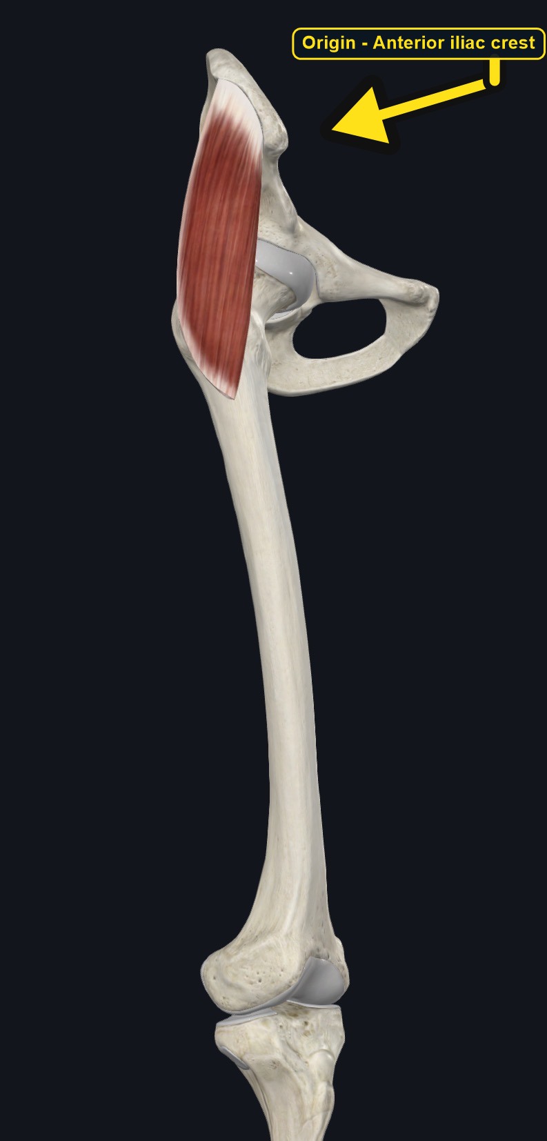
| Origin | The anterior part of the outer lip of the iliac crest, near the anterior superior iliac spine. |
|---|
Section 03 of 20
Insertion
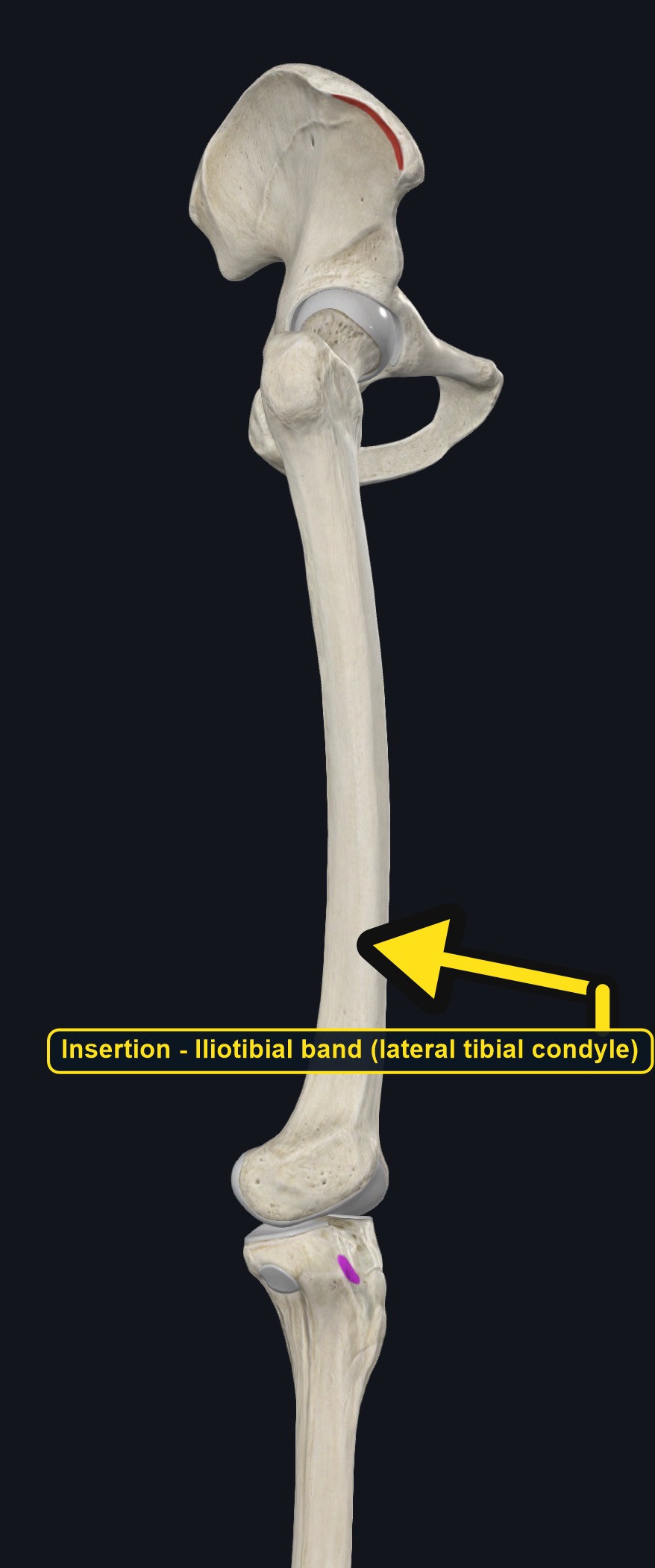
| Insertion | The iliotibial band, which continues to the lateral condyle of the tibia (Gerdy's tubercle). |
|---|
Section 04 of 20
Actions
No dedicated action video is currently available. This movement is explained below and video will be added when verified source media becomes available.
| Primary action | Hip flexion and abduction. |
|---|---|
| Secondary actions | Medial rotation of the thigh and tensioning of the iliotibial band for pelvic and knee stability. |
| Movement at the joint | Hip joint, with an indirect effect at the knee through the iliotibial band. |
| Heads / fiber groups | Single small muscle feeding into the iliotibial band. |
| Functional examples | Walking and running (stance phase); cycling; side-stepping; getting out of a car. |
Hip flexion
Helps flex the thigh at the hip.
Hip abduction
Draws the thigh away from the midline.
Medial rotation of the thigh
Turns the thigh inward.
Knee and pelvic stabilization
Tenses the iliotibial band, helping steady the outer knee and the pelvis during single-leg stance.
Section 05 of 20
Nerve Innervation
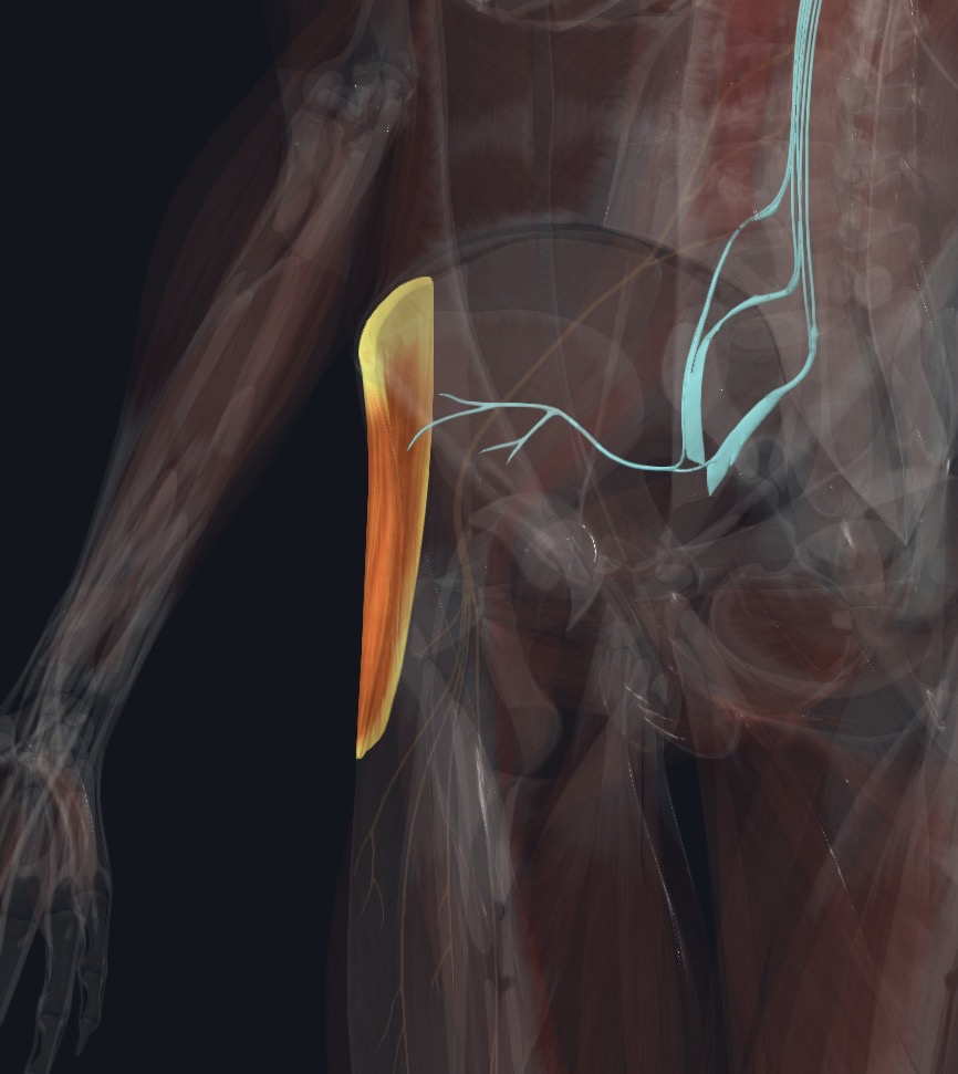
| Nerve | Superior gluteal nerve, roots L4–S1. |
|---|
The superior gluteal nerve also supplies gluteus medius and minimus, so weakness affecting all three muscles may be associated with a pelvic drop on the opposite side when walking (a Trendelenburg pattern), which may reflect a nerve or hip joint problem.
Section 06 of 20
Blood Supply
| Blood | The ascending branch of the lateral circumflex femoral artery, with contributions from the superior gluteal artery. |
|---|
Section 07 of 20
Pictures and Visual Anatomy
The pictures for this muscle are shown with the Origin, Insertion, Nerve Innervation, and other sections where they teach best.
Section 08 of 20
Functional Movement
Tensor fasciae latae is active in walking, running, and cycling, especially in the stance phase when it helps keep the pelvis level and the knee stable. It also works during activities like side-stepping and getting out of a car.
Section 09 of 20
Synergists and Antagonists
Synergists: gluteus medius and minimus (abduction and medial rotation), iliopsoas, rectus femoris, and sartorius (hip flexion), and gluteus maximus for tensioning the iliotibial band. Antagonists: gluteus maximus and the hamstrings (hip extension), the adductors (hip adduction), and the lateral rotators (lateral rotation).
Section 10 of 20
Palpation and Location
Client position and technique
Position the client side-lying with the underneath leg bent and the upper leg straight. Locate the front of the iliac crest, then find the short, thick muscle belly just below it and in front of gluteus medius. Ask the client to gently lift the leg forward and out (flexion with abduction) to feel it tighten, and follow the iliotibial band as it continues down the outer thigh.
Protect nearby structures
Use moderate, broad pressure over the muscle belly and lighter pressure along the iliotibial band, which is fascia rather than muscle and can feel tender when tight. Stop if the client reports sharp pain or symptoms that spread down the leg.
Section 11 of 20
Massage Therapy Relevance
Tensor fasciae latae and the iliotibial band are frequently involved in outer hip and outer knee pain in runners and cyclists. Because iliotibial band pain has several possible sources, careful assessment matters.
Section 12 of 20
Dysfunction: What Happens When Movement Goes Wrong
Changes in how a muscle works can change how a joint moves. The points below describe possibilities that may be associated with altered muscle function; they are not diagnoses, and one muscle is never assumed to be the sole cause of a problem.
When the muscle is weak
Weakness may be associated with reduced pelvic control on one-leg stance and may contribute to a Trendelenburg-type gait pattern.
When it is tight or shortened
Tightness may be associated with anterior pelvic tilt, limited hip extension and adduction, and tension along the iliotibial band.
When it is overused
Repeated running, cycling, and hip flexion may overload the muscle and the iliotibial band and may contribute to outer hip or knee pain.
When it is strained
A strain may cause outer hip pain with resisted hip flexion or abduction; hip joint and trochanteric bursa problems can look similar.
When it is inhibited or others compensate
Reduced activation may be associated with more work by gluteus medius and altered pelvic control.
When it does not coordinate well with synergists and antagonists
Poor coordination with gluteus medius, gluteus maximus, and the hip flexors may alter pelvic and knee control and may be associated with hip or knee symptoms.
Section 13 of 20
Conditions, Pathologies, and Pain Relationships
Iliotibial band syndrome
Repetitive knee bending, as in running or cycling, may irritate the iliotibial band near the outer knee, causing pain with activity.
Greater trochanteric pain syndrome
Tightness in TFL and the iliotibial band may add tension over the greater trochanter and may be associated with outer hip pain.
Hip flexor tightness
A short TFL may be associated with anterior pelvic tilt and reduced hip extension, sometimes contributing to low back tension.
Differential considerations
Hip joint problems, lateral meniscus injury, lumbar referral, and lateral collateral ligament injury of the knee can all cause similar outer hip or knee pain.
Section 14 of 20
Example Clinical Relationship
A runner reports a sharp pain on the outside of the knee that appears after a mile or two and eases with rest. Tensor fasciae latae and the iliotibial band may be involved, and the pattern may be associated with iliotibial band syndrome, but a lateral meniscus or ligament injury can look similar. Gentle work on the TFL muscle belly may support comfort, while a knee that gives way and swells, or sudden pain after a fall, needs referral.
Section 15 of 20
Massage and Treatment Approaches
For healthy, non-acute tissue, position the client side-lying with pillows for support. Begin with broad effleurage over the outer hip and thigh, then use gentle kneading of the TFL muscle belly and lighter, longer strokes along the iliotibial band, staying within comfortable pressure.
| Movement | Use gentle hip extension and adduction stretches within comfort; do not force range. |
|---|---|
| Regional work | Consider gluteus medius and maximus, the outer quadriceps, and the lateral knee structures when assessment supports it. |
| Avoid | Deep pressure directly over the greater trochanter or the lateral knee if acutely tender, or at any point that reproduces sharp or radiating pain. |
Section 16 of 20
Precautions and Contraindications
Precautions and contraindications
- Acute strain, recent hip or knee injury or surgery, or suspected fracture.
- Use caution with known greater trochanteric bursitis or iliotibial band syndrome; avoid aggressive friction directly over an acutely inflamed area.
- Use caution with anticoagulant use or easy bruising.
- Stop if pressure causes sharp pain or symptoms that spread down the leg.
Section 17 of 20
Red Flags and When to Refer
Red flags and referral
- Sudden hip or knee pain after a fall, or inability to bear weight.
- Hip or thigh pain with fever, night pain, or unexplained weight loss.
- Progressive leg weakness, numbness, or a knee that gives way repeatedly and swells.
- Signs of a blood clot: a swollen, warm, red, or painful leg on one side.
Section 18 of 20
Study Summary
Attachments
Origin: anterior iliac crest. Insertion: iliotibial band to the lateral tibial condyle.
Actions
Hip flexion, abduction, and medial rotation; stabilizes the pelvis and knee via the iliotibial band.
Supply
Superior gluteal nerve (L4–S1); ascending branch of the lateral circumflex femoral artery.
Section 19 of 20
Instructor Narration
Open narration transcript
Tensor fasciae latae is a short, thick muscle at the front of the outer hip that feeds into the iliotibial band.
It arises from the front of the iliac crest and inserts into the iliotibial band, which continues to the lateral tibial condyle at Gerdy's tubercle.
It flexes, abducts, and medially rotates the hip, and tenses the iliotibial band to help stabilize the pelvis and knee. Gluteus medius and minimus help it, and gluteus maximus and the hamstrings oppose it.
The superior gluteal nerve supplies it, from L4 to S1, and the ascending branch of the lateral circumflex femoral artery provides its blood.
Palpate with the client side-lying, just below the front of the iliac crest and in front of gluteus medius, and follow the iliotibial band down the outer thigh. Use lighter pressure along the band itself.
Iliotibial band syndrome, greater trochanteric pain, and hip flexor tightness are common, and hip joint, knee, and lumbar problems can overlap. Refer sudden hip or knee pain after a fall, hip pain with fever, or a knee that gives way and swells.
Remember: anterior iliac crest to the iliotibial band and Gerdy's tubercle; hip flexion, abduction, and medial rotation; superior gluteal nerve.
Section 20 of 20