Section 01 of 20
Introduction
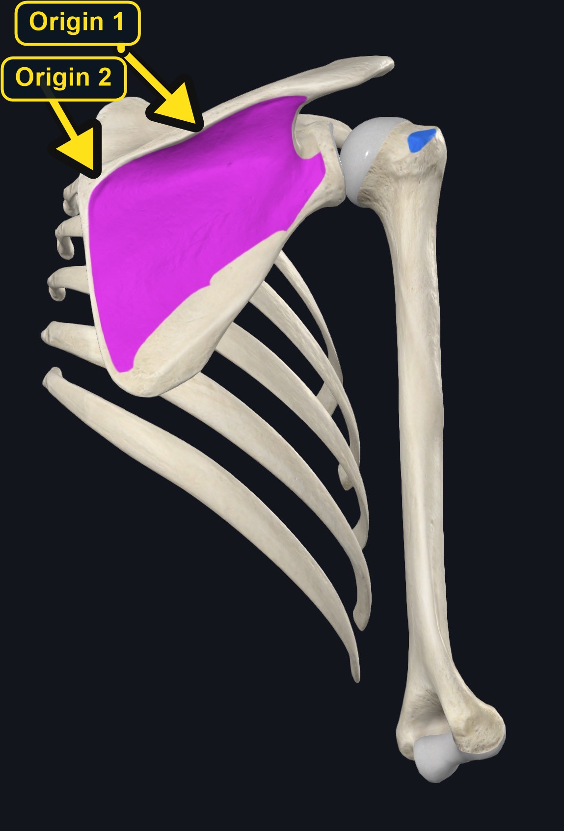
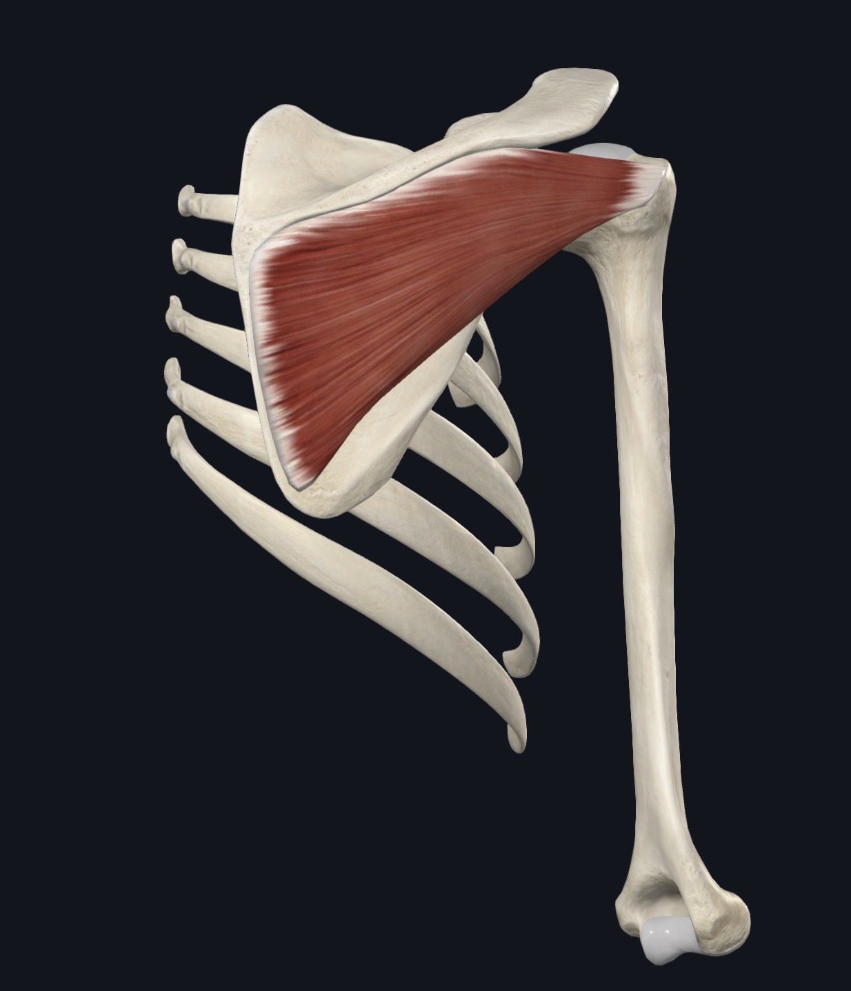
Subscapularis is the largest and most powerful of the four rotator cuff muscles. Unlike the other three (supraspinatus, infraspinatus, and teres minor), which lie on the back of the scapula, subscapularis occupies the entire front (subscapular fossa) surface of the shoulder blade, sandwiched between the scapula and the rib cage.
As the primary internal rotator of the shoulder and a major contributor to anterior joint stability, subscapularis plays an essential role in resisting excessive external rotation and anterior translation of the humeral head — the same direction of force involved in most anterior shoulder dislocations.
For massage therapists, subscapularis is a genuinely challenging muscle to access directly because of its deep, anterior position against the rib cage, but understanding its anatomy and function is still important for a complete picture of shoulder mechanics, rotator cuff pathology, and conditions like adhesive capsulitis.
Section 02 of 20
Origin
| Origin | Subscapular fossa — the broad, shallow concave surface on the anterior (rib-cage-facing) surface of the scapula. |
|---|
Section 03 of 20
Insertion
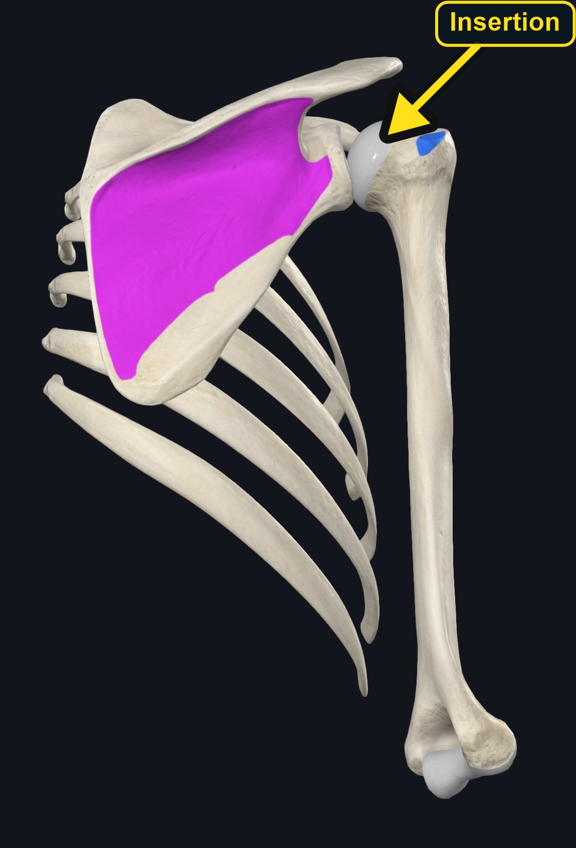
| Insertion | Lesser tubercle of the humerus, with fibers also blending into the anterior shoulder joint capsule. |
|---|
Section 04 of 20
Actions
| Primary action | Internal (medial) rotation of the shoulder. |
|---|---|
| Secondary actions | Stabilizes the humeral head anteriorly; assists adduction and, with the arm elevated, extension. |
| Movement at the joint | Glenohumeral (shoulder) joint. |
| Heads / fiber groups | Upper and lower parts, supplied by the upper and lower subscapular nerves. |
| Functional examples | Placing the hand behind the back; throwing; swimming; keeping the shoulder stable when lifting. |
Primary actions
- Internal (medial) rotation of the shoulder — subscapularis is generally considered the primary and most powerful internal rotator among the shoulder muscles.
Secondary / functional role
- As part of the rotator cuff, contributes significantly to dynamic anterior stabilization of the glenohumeral joint, helping to resist excessive anterior translation and external rotation of the humeral head — a role especially important near the end range of shoulder external rotation and abduction (such as the cocking phase of a throw).
Section 05 of 20
Nerve Innervation
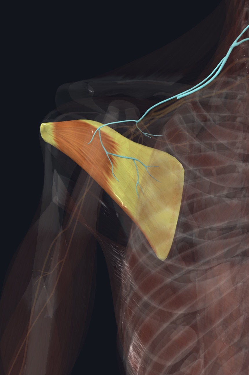
| Nerve | Upper and lower subscapular nerves |
|---|---|
| Roots | C5, C6 (upper subscapular nerve primarily C5-C6; lower subscapular nerve, which also supplies teres major, primarily C5-C7 depending on source) |
| Relationship | Subscapularis is unusual among the rotator cuff muscles in receiving dual innervation from two separate named nerves — the upper and lower subscapular nerves — both branches of the posterior cord of the brachial plexus. |
Section 06 of 20
Blood Supply
Subscapularis is supplied primarily by the subscapular artery and its branches, along with contribution from the circumflex scapular artery.
Section 07 of 20
Pictures and Visual Anatomy
Section 08 of 20
Functional Movement
Subscapularis turns the arm inward and holds the front of the humeral head steady in the shoulder socket. It is active when placing the hand behind the back, throwing, swimming, and lifting the arm while keeping the shoulder stable.
Section 09 of 20
Synergists and Antagonists
Synergists
Pectoralis major, latissimus dorsi, and teres major (internal rotation).
Antagonists
Infraspinatus and teres minor (external rotation).
Section 10 of 20
Palpation and Location
Client position
Client supine, arm relaxed at the side or gently supported in slight abduction, to allow limited access to the axillary border of the scapula.
Landmarks and technique
- Direct palpation of subscapularis is inherently limited because the muscle lies against the rib cage on the front surface of the scapula, largely inaccessible through normal soft tissue approaches.
- The muscle can be partially accessed along the lateral border of the scapula within the axilla, with the client's arm relaxed and gently supported, approaching gradually and gently from the axillary region with the fingers curving slightly toward the front surface of the scapula — this requires a light, gradual, feedback-guided approach given the sensitivity of the axilla.
- Resisted internal rotation (with the elbow tucked at the side, forearm pressing inward against light resistance) can help confirm general muscle activity even where direct palpation is limited.
- Given the depth and sensitivity of this region, and the proximity of the axillary neurovascular bundle, this is an advanced palpation area that should only be approached with appropriate training and always guided closely by client comfort and consent.
Section 11 of 20
Massage Therapy Relevance
Subscapularis is particularly relevant in discussions of adhesive capsulitis ("frozen shoulder"), where restriction of the anterior joint capsule and surrounding structures, including subscapularis, is commonly associated with significant loss of external rotation range of motion.
Clients with a history of shoulder instability, overhead throwing sports, or general anterior shoulder tightness related to a rounded-shoulder posture may also show increased tone or shortening in subscapularis, given its role as a primary internal rotator alongside other anteriorly-positioned muscles like pectoralis major.
Because direct access to this muscle is limited and requires advanced technique, general massage therapy approaches often address subscapularis indirectly, through work on the surrounding shoulder girdle and gentle range-of-motion techniques, rather than through direct deep tissue contact.
Section 12 of 20
Dysfunction: What Happens When Movement Goes Wrong
Changes in how a muscle works can change how a joint moves. The points below describe possibilities that may be associated with altered muscle function; they are not diagnoses, and one muscle is never assumed to be the sole cause of a problem.
When the muscle is weak
Weakness may reduce internal rotation strength and may be associated with anterior instability and a positive lift-off type test.
When it is tight or shortened
Tightness may be associated with limited external rotation and may contribute to a stiff shoulder, sometimes in a picture that overlaps with adhesive capsulitis.
When it is overused
Throwing, swimming, and repetitive overhead work may load the muscle and may contribute to deep front-of-shoulder ache.
When it is strained
A strain or tendon problem may cause anterior shoulder pain with resisted internal rotation; rotator cuff tears and biceps tendon problems can look similar.
When it is inhibited or others compensate
Reduced activation may be associated with less anterior shoulder stability and compensation from pectoralis major and latissimus dorsi.
When it does not coordinate well with synergists and antagonists
Poor balance with infraspinatus and the other rotator cuff muscles may alter humeral head control and may be associated with shoulder pain.
Section 13 of 20
Conditions, Pathologies, and Pain Relationships
Subscapularis tendinopathy / tear
What it is: Irritation, degenerative change, or structural tearing of the subscapularis tendon, less common than supraspinatus involvement but clinically significant when present.
Why the muscle may be involved: Repetitive internal rotation loading, or trauma involving forced external rotation that can overstretch or tear the tendon.
Typical symptoms: Anterior shoulder pain, weakness with resisted internal rotation, and in some clinical assessments, an increased passive external rotation range due to loss of the muscle's normal restraining function.
Similar-presenting conditions: Biceps tendon pathology (the long head tendon runs near subscapularis at the bicipital groove), other rotator cuff involvement, anterior shoulder instability.
Refer when: Suspected tear (notable weakness, especially after trauma) warrants medical/orthopedic evaluation.
Adhesive capsulitis (frozen shoulder) — general association
What it is: A condition involving progressive stiffening and pain of the shoulder joint capsule, typically progressing through recognized phases (freezing, frozen, thawing).
Why the muscle may be involved: The anterior capsule, closely associated with subscapularis, is commonly involved in the restrictive process, and loss of external rotation is a hallmark clinical sign.
Typical symptoms: Progressive shoulder stiffness and pain, often with significant loss of both active and passive range of motion in multiple directions.
Similar-presenting conditions: Rotator cuff pathology, osteoarthritis of the glenohumeral joint, other causes of shoulder stiffness.
Refer when: Adhesive capsulitis is a medical diagnosis; clients with significant, progressive shoulder stiffness should be evaluated by a healthcare provider, though gentle supportive massage therapy may be appropriate within scope once a client has appropriate medical guidance.
Anterior shoulder instability (general association)
What it is: A tendency toward excessive anterior translation or dislocation of the humeral head.
Why the muscle may be involved: Subscapularis is a key dynamic anterior stabilizer; its function is relevant to overall anterior joint stability, though instability itself usually involves other structures (capsule, labrum) as well.
Typical symptoms: A sensation of the shoulder feeling loose, apprehension with certain arm positions (particularly abduction combined with external rotation), or history of dislocation/subluxation.
Similar-presenting conditions: Labral tears, general ligamentous laxity, rotator cuff dysfunction.
Refer when: Any suspected instability or history of dislocation warrants orthopedic evaluation; this is outside massage therapy scope to assess or treat directly.
Section 14 of 20
Example Clinical Relationship
Why external rotation loss is a hallmark sign
Because subscapularis is the primary internal rotator and lies against the anterior joint capsule, conditions that restrict the anterior capsule — most notably adhesive capsulitis — often present with a characteristic and disproportionate loss of external rotation range of motion, since the tight anterior structures resist the stretch required to rotate the arm outward. This is one reason external rotation range of motion is a commonly referenced clinical sign when adhesive capsulitis is suspected.
Massage therapists should understand this relationship as useful background for recognizing a pattern that may warrant medical referral (progressive, significant loss of shoulder motion in multiple directions, often with a gradual, non-traumatic onset), rather than as a basis for diagnosing frozen shoulder themselves. Once a client has appropriate medical guidance, gentle range-of-motion-supportive techniques and general soft tissue work on the accessible surrounding shoulder girdle musculature may be considered as one part of a broader care plan, always within massage therapy scope and working alongside, not instead of, appropriate medical management.
Section 15 of 20
Massage and Treatment Approaches
The following approaches may be considered for a generally healthy client with no contraindications present.
Broad warming strokes (surrounding region)
Why: Since subscapularis itself is not directly broadly accessible, general warming of the surrounding shoulder girdle and axillary border can support overall comfort and assessment.
Direction/location: Broad strokes across the anterior shoulder, chest, and lateral scapular border, respecting draping and consent.
Pressure: Light.
Gentle, gradual axillary-border access (advanced technique)
Why: Some trained therapists use a careful, gradual approach along the lateral scapular border within the axilla to access a portion of subscapularis.
Direction/location: Along the axillary border of the scapula, with the client's arm relaxed and supported.
Pressure: Very light to start, increasing only gradually and only within appropriate training and clear client consent; this is an advanced, sensitive-area technique and should not be attempted without proper training.
Avoid: Any pressure that produces numbness, tingling, or sharp pain — stop immediately.
Gentle passive/active range of motion
Why: Gentle, pain-free range-of-motion work can support general shoulder mobility and comfort, particularly useful for clients working on regaining motion as part of a broader care plan (e.g., alongside physical therapy for frozen shoulder).
Direction/location: Gentle passive external rotation, staying strictly within pain-free range.
Pressure: Light; never force through resistance or pain.
Myofascial techniques (surrounding tissue)
Why: May help address general fascial restriction in the accessible surrounding shoulder girdle.
Direction/location: Slow, broad contact across the anterior shoulder and chest.
Pressure: Light, sustained.
Section 16 of 20
Precautions and Contraindications
Use caution or avoid the area when any of the following are present
- Acute shoulder injury or suspected rotator cuff tear.
- Suspected shoulder instability or history of dislocation without medical clearance.
- Recent shoulder surgery without medical clearance for massage.
- Signs of acute inflammation or infection.
- Significant, progressive, or severe loss of shoulder motion of unclear cause — appropriate for medical evaluation before extensive manual therapy.
Vulnerable structures — avoid firm direct pressure
- The axilla and its neurovascular contents — any direct access technique in this region should be gentle, gradual, and performed only with appropriate training.
Section 17 of 20
Red Flags and When to Refer
Postpone massage and refer for medical evaluation if a client reports or shows:
- Significant or progressive loss of shoulder motion, especially with a non-traumatic, gradual onset.
- New numbness, tingling, or weakness in the arm or hand.
- A sensation of shoulder instability or apprehension with certain arm positions, or history of dislocation.
- Signs of infection (fever, redness, warmth, swelling).
- Unexplained, severe, or worsening pain not clearly related to a specific movement or activity.
- Recent significant trauma to the shoulder.
Section 18 of 20
Study Summary
Subscapular fossa (anterior surface of the scapula).
Lesser tubercle of the humerus.
Primary internal rotation of the shoulder; major anterior joint stabilizer.
Upper and lower subscapular nerves, primarily C5-C6 with C7 contribution described in some references.
Largest rotator cuff muscle; limited direct access; relevant to adhesive capsulitis and anterior shoulder stability discussions.
Section 19 of 20
Instructor Narration
Open narration script (readable / for audio conversion)
Section 20 of 20