Section 01 of 20
Introduction
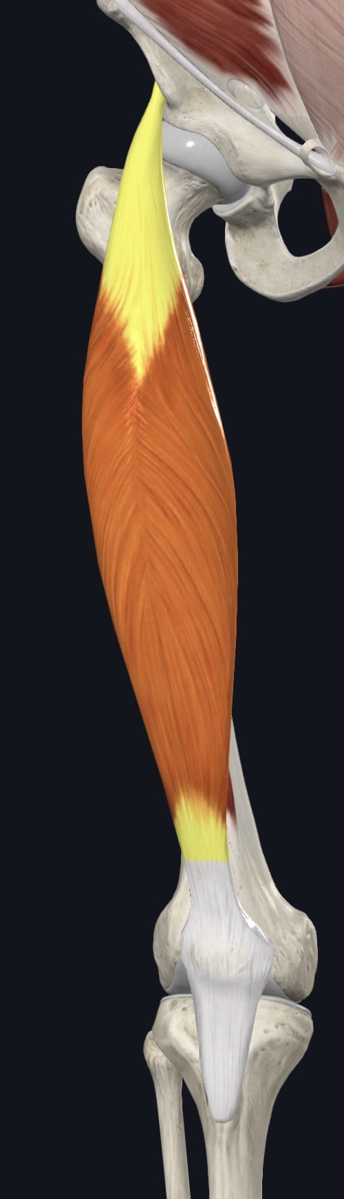
Rectus femoris is a spindle-shaped muscle in the center of the front of the thigh, lying over the vastus intermedius. It is one of the four muscles of the quadriceps group, and it is the only one that crosses the hip joint as well as the knee.
Its two-joint design means it works best when the hip and knee move together, for example when kicking a ball. It joins the other three quadriceps muscles in the quadriceps tendon, which surrounds the patella and continues as the patellar ligament to the tibia.
Section 02 of 20
Origin
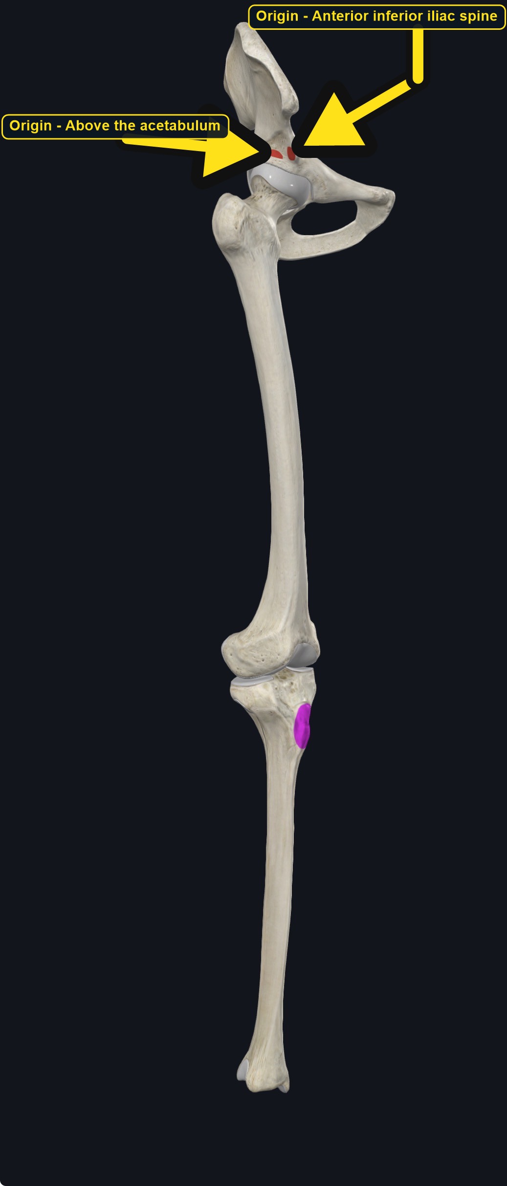
| Origin | Straight head: the anterior inferior iliac spine (AIIS). Reflected head: the groove just above the acetabulum (the hip socket). |
|---|
Section 03 of 20
Insertion
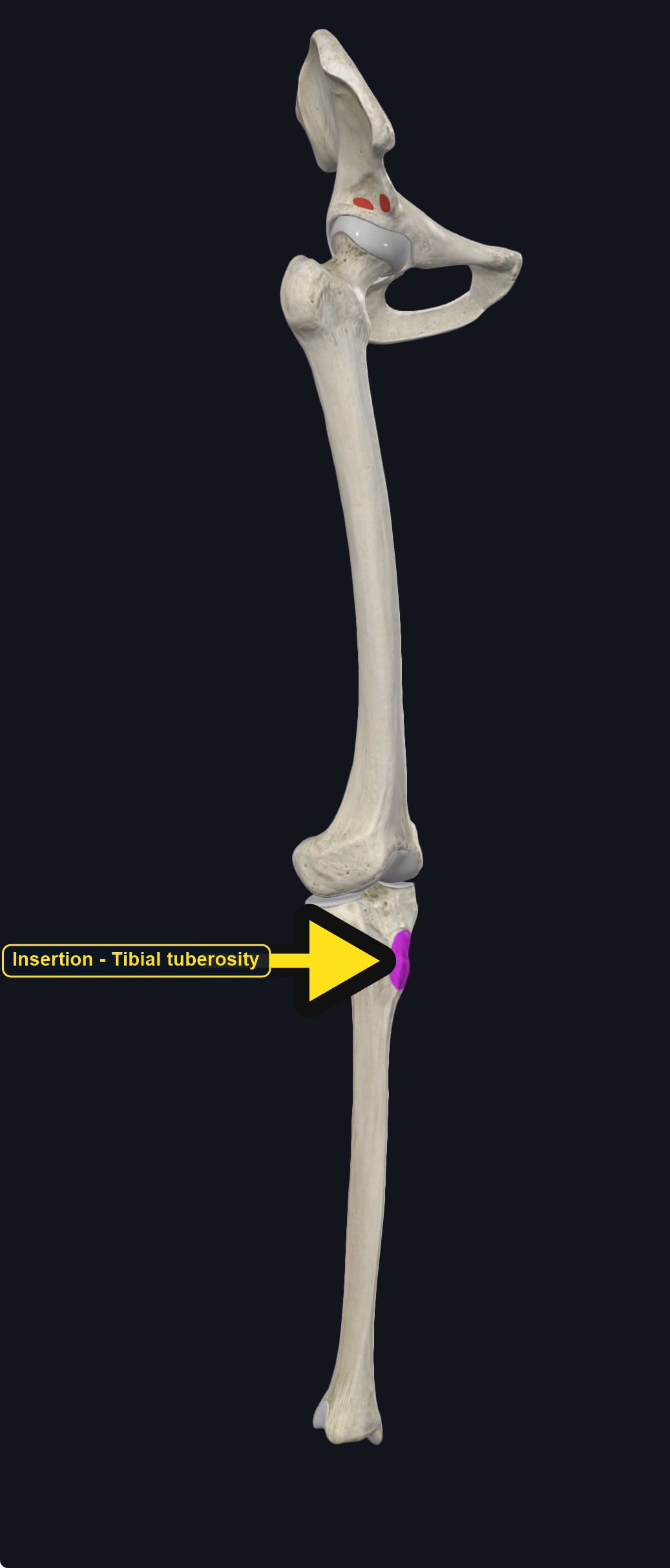
| Insertion | The base of the patella through the quadriceps tendon, and then the tibial tuberosity through the patellar ligament. |
|---|
Section 04 of 20
Actions
No dedicated action video is currently available. This movement is explained below and video will be added when verified source media becomes available.
| Primary action | Knee extension. |
|---|---|
| Secondary actions | Hip flexion, and stabilization of the hip and knee. |
| Movement at the joint | Knee joint and hip joint. |
| Heads / fiber groups | Two heads at the origin (straight and reflected) with one muscle belly. |
| Functional examples | Kicking; sprinting; jumping; climbing stairs; rising from a chair; cycling. |
Knee extension
Straightens the knee together with the three vasti muscles.
Hip flexion
Lifts the thigh forward at the hip, the only quadriceps head able to do so.
Pelvic control
Helps tilt the pelvis forward when the leg is fixed and stabilizes the hip and knee during movement.
Section 05 of 20
Nerve Innervation
| Nerve | Femoral nerve, roots L2–L4. |
|---|
The femoral nerve and vessels run close to the top of the thigh. Weakness of knee extension with a knee that gives way may reflect a femoral nerve problem, not only muscle weakness.
Section 06 of 20
Blood Supply
| Blood | The descending branch of the lateral circumflex femoral artery, with branches of the femoral artery. |
|---|
Section 07 of 20
Pictures and Visual Anatomy
Section 08 of 20
Functional Movement
Rectus femoris is used when kicking, sprinting, jumping, climbing stairs, rising from a chair, and cycling. Because it crosses two joints, it is especially active in the swing phase of walking and running when the hip flexes and the knee extends together.
Section 09 of 20
Synergists and Antagonists
Synergists: vastus lateralis, vastus medialis, and vastus intermedius (knee extension), and iliopsoas, sartorius, tensor fasciae latae, and pectineus (hip flexion). Antagonists: the hamstrings (knee flexion and hip extension) and gluteus maximus (hip extension).
Section 10 of 20
Palpation and Location
Client position and technique
Position the client supine with the knee slightly supported. Locate the anterior inferior iliac spine just below the anterior superior iliac spine, then follow the muscle down the center of the front of the thigh toward the patella. Ask for a straight leg raise or a gentle knee extension to feel the muscle tighten.
Protect nearby structures
The femoral triangle at the top of the thigh contains the femoral artery, vein, and nerve. Stay below the inguinal crease, avoid pressure on a pulse, and stop if the client reports tingling, numbness, or sharp pain.
Section 11 of 20
Massage Therapy Relevance
Rectus femoris is a common site of tightness in people who sit a lot, and of strain in runners, sprinters, and kickers. It is often assessed with hip flexor length, knee pain, and anterior pelvic tilt, but many joint, tendon, and nerve conditions can cause similar symptoms.
Section 12 of 20
Dysfunction: What Happens When Movement Goes Wrong
Changes in how a muscle works can change how a joint moves. The points below describe possibilities that may be associated with altered muscle function; they are not diagnoses, and one muscle is never assumed to be the sole cause of a problem.
When the muscle is weak
Weakness may reduce knee extension strength and may be associated with a knee that gives way and more work by the vasti and hip flexors.
When it is tight or shortened
Tightness may be associated with anterior pelvic tilt, limited hip extension and knee flexion, and pain at the front of the hip or knee.
When it is overused
Sprinting, kicking, and repeated jumping may overload the muscle and tendon and may contribute to thigh or knee pain.
When it is strained
A strain may cause sudden front-of-thigh or hip pain with kicking or hip flexion; hip joint problems can look similar.
When it is inhibited or others compensate
Reduced activation may be associated with more work by the iliopsoas and vasti and altered knee tracking.
When it does not coordinate well with synergists and antagonists
Poor coordination with the vasti, hamstrings, and gluteal muscles may alter knee and pelvic control and may be associated with knee or hip symptoms.
Section 13 of 20
Conditions, Pathologies, and Pain Relationships
Rectus femoris strain
Sprinting and kicking can strain the muscle or its upper tendon, causing sudden pain at the front of the hip or thigh and difficulty flexing the hip.
Quadriceps tendinopathy and patellofemoral pain
Overload of the quadriceps tendon, or altered pull on the patella, may be associated with pain above or around the kneecap.
Contusion and myositis ossificans
A direct blow to the thigh can cause a deep bruise; if it does not settle, bone-like tissue can form in the muscle and needs medical assessment.
Differential considerations
Hip joint problems, femoral nerve entrapment, referred lumbar pain, and stress injuries can all cause front-of-thigh and knee pain.
Section 14 of 20
Example Clinical Relationship
A sprinter reports a sudden sharp pain at the front of the hip while accelerating, and now feels a deep ache with hip flexion. Rectus femoris may be involved, and the pattern may be associated with a strain of its upper tendon, but hip joint and other muscle problems can look similar. Gentle work on nearby tissue may support comfort after assessment, while a sudden gap, severe pain, or a swollen warm leg needs referral.
Section 15 of 20
Massage and Treatment Approaches
For healthy, non-acute tissue, position the client supine with the knee supported. Begin with broad effleurage from the knee toward the hip, then use kneading, compression, and slow longitudinal strokes along the muscle, adding gentle knee flexion and hip extension stretches within comfort.
| Movement | Use gentle passive knee flexion with the hip extended (or side-lying quad stretch) within comfort; do not force stretch. |
|---|---|
| Regional work | Consider the other quadriceps muscles, the iliopsoas, tensor fasciae latae, sartorius, and the iliotibial band when assessment supports it. |
| Avoid | Deep pressure in the femoral triangle, over an acute strain or a deep bruise, or at any point that reproduces leg tingling. |
Section 16 of 20
Precautions and Contraindications
Precautions and contraindications
- Acute strain or tear, a recent deep bruise, or recent hip, thigh, or knee injury or surgery.
- Use caution with anticoagulant use, easy bruising, or a history of thigh trauma with a hard lump.
- Avoid deep pressure over the femoral triangle and over varicose veins.
- Stop if pressure causes sharp pain, numbness, or tingling.
Section 17 of 20
Red Flags and When to Refer
Red flags and referral
- A hard, painful lump in the thigh after an injury that is not settling.
- A swollen, warm, red, or painful leg, especially on one side, or sudden shortness of breath (possible blood clot).
- Sudden severe pain and a bulge or gap in the muscle after sprinting or kicking, or inability to flex the hip.
- Thigh pain with fever, night pain, or unexplained weight loss.
Section 18 of 20
Study Summary
Attachments
Origin: AIIS and the groove above the acetabulum. Insertion: patella, then the tibial tuberosity.
Actions
Knee extension and hip flexion.
Supply
Femoral nerve (L2–L4); lateral circumflex femoral artery.
Section 19 of 20
Instructor Narration
Open narration transcript
Rectus femoris is the central muscle of the front of the thigh and the only quadriceps muscle that crosses the hip.
It arises from the anterior inferior iliac spine and the groove above the acetabulum, and inserts on the patella through the quadriceps tendon, and then on the tibial tuberosity through the patellar ligament.
It extends the knee and flexes the hip. The three vasti extend the knee with it, and the iliopsoas and sartorius help flex the hip. The hamstrings and gluteus maximus oppose it.
The femoral nerve supplies it, from L2 to L4, and the lateral circumflex femoral artery provides its blood.
Palpate with the client supine, starting at the anterior inferior iliac spine and following the muscle down the thigh with a straight leg raise. Stay below the inguinal crease and off any pulse.
Strain, tendinopathy, contusion with possible bone formation, and hip and nerve problems can overlap. Refer a hard lump after injury, a swollen warm leg, a sudden gap with severe pain, or thigh pain with fever.
Remember: AIIS and above the acetabulum to the patella and tibial tuberosity; knee extension and hip flexion; femoral nerve.
Section 20 of 20