Section 01 of 20
Introduction
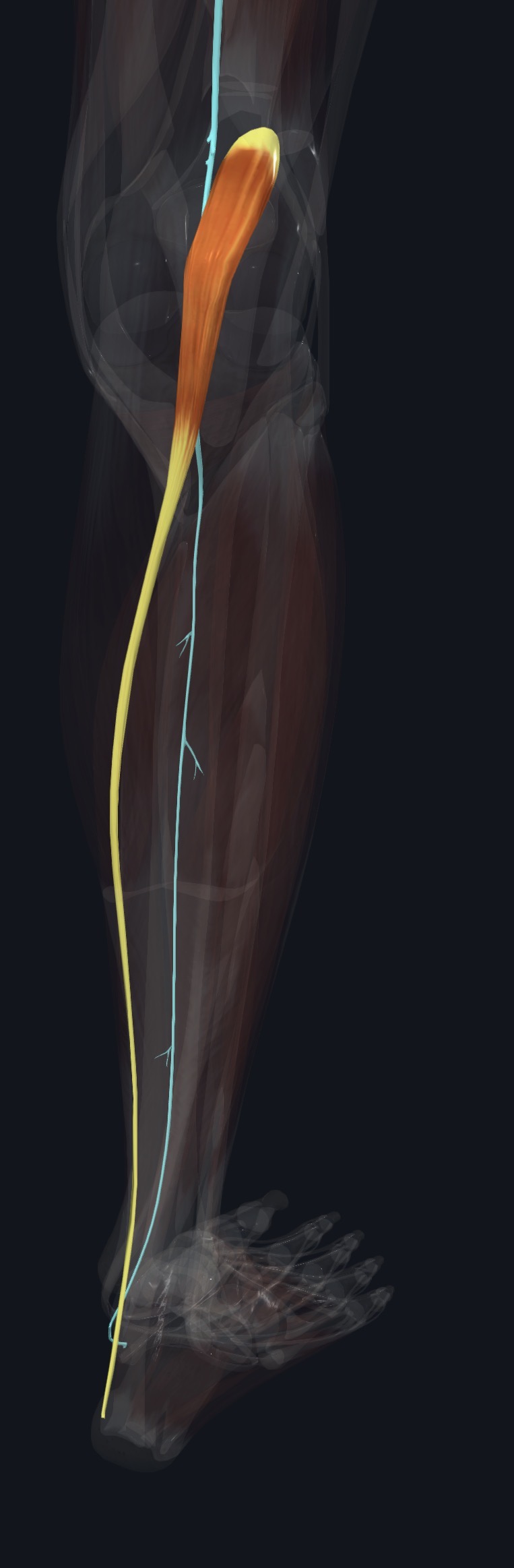
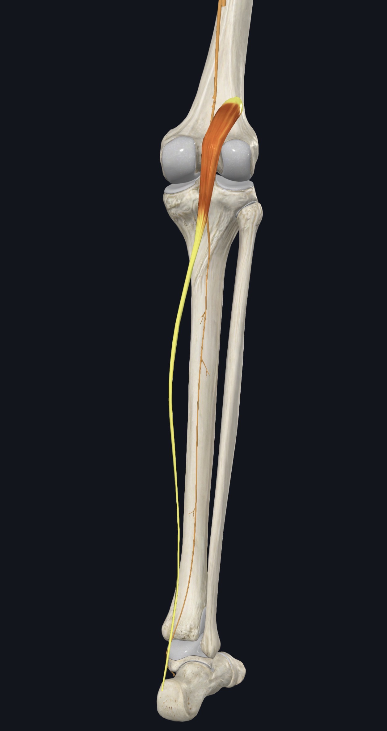
Plantaris is a small muscle with a short belly, about the size of a finger, high in the back of the leg, and an extremely long, thin tendon. The belly lies behind the knee between the lateral head of gastrocnemius and the soleus, and the tendon runs down between them to reach the heel.
It is absent in a small proportion of people (commonly reported around 7 to 10 percent), and this causes no noticeable loss of function. It is often described as a helper of gastrocnemius rather than a major mover.
Section 02 of 20
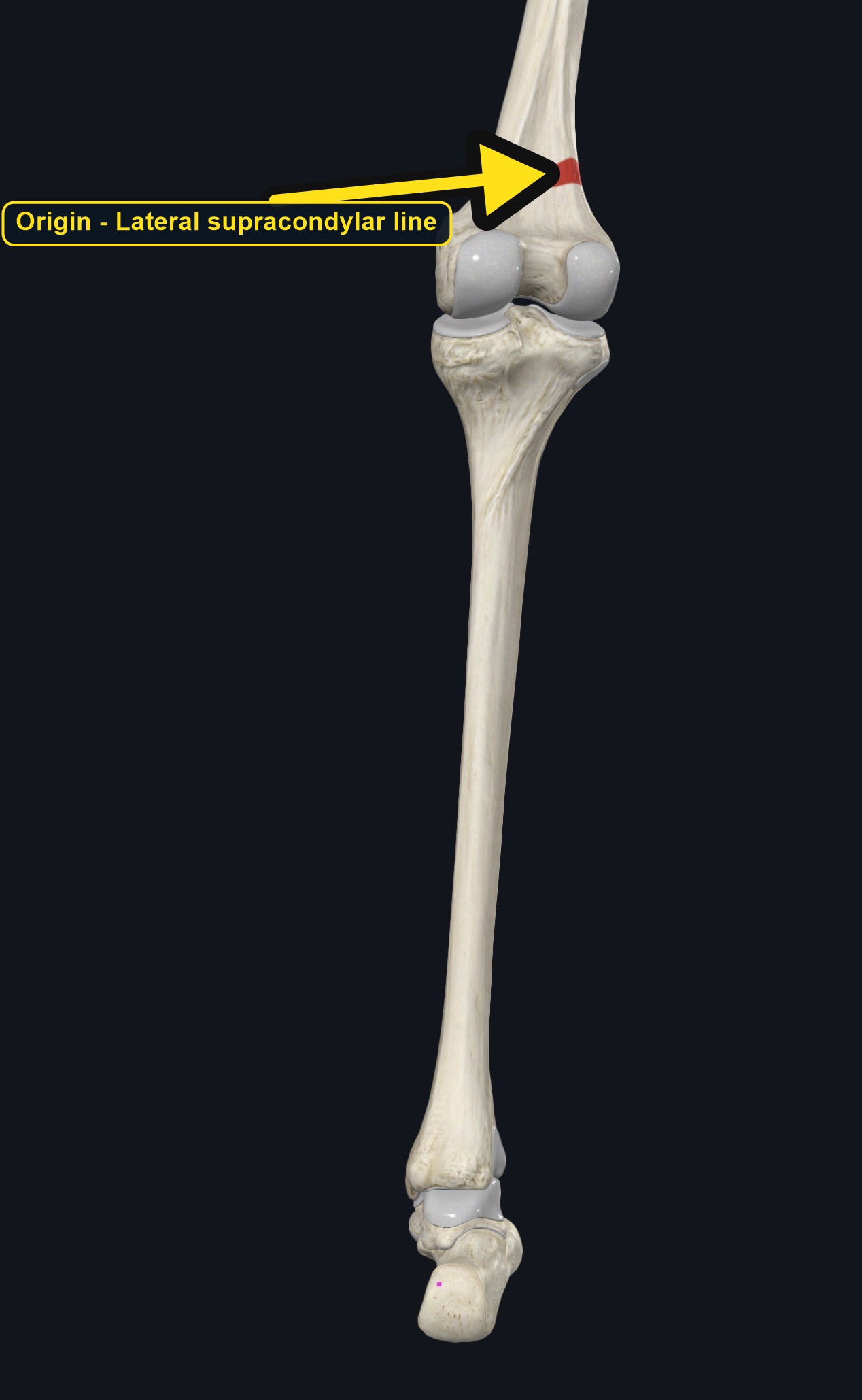
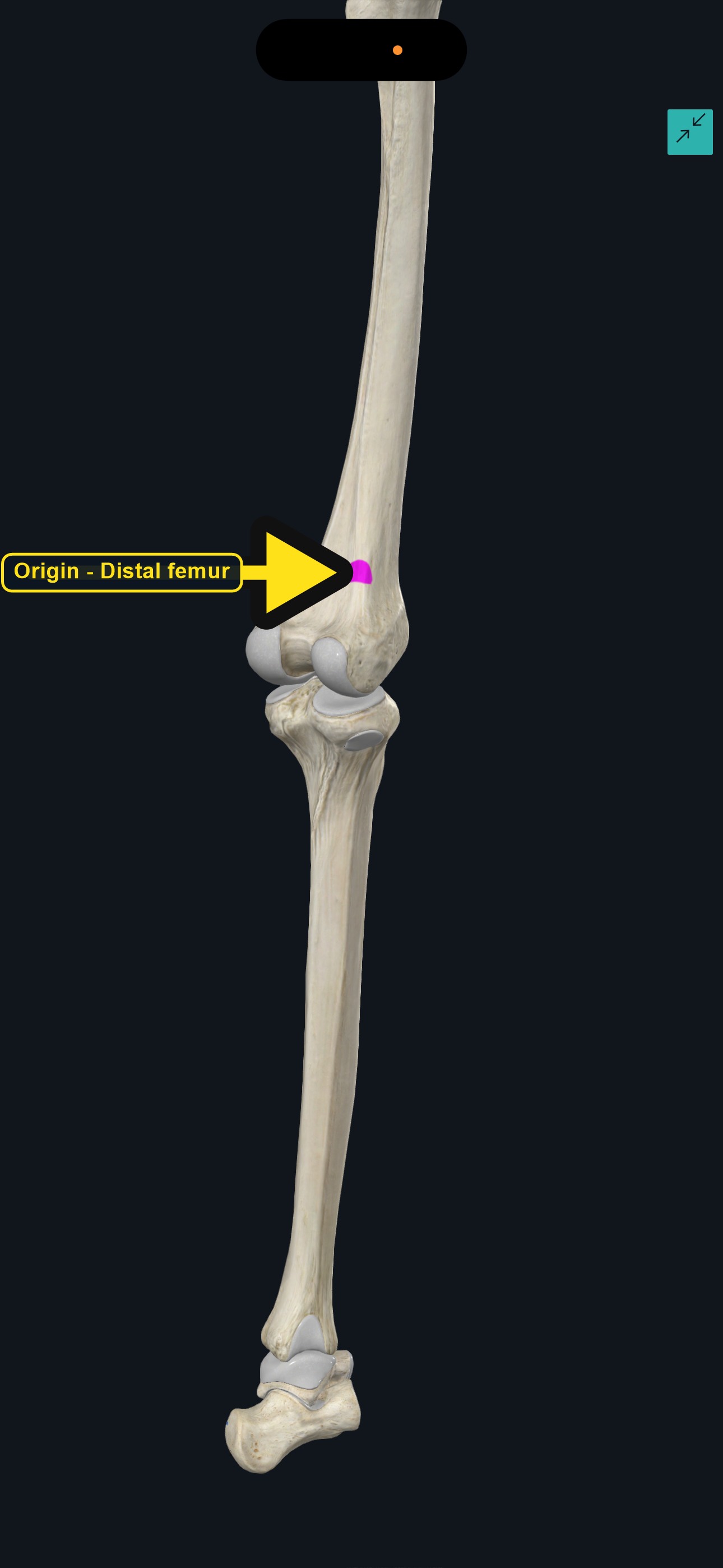
Origin
| Origin | Inferior part of the lateral supracondylar line of the femur, just above the lateral condyle, and the oblique popliteal ligament of the knee. |
|---|
Section 03 of 20
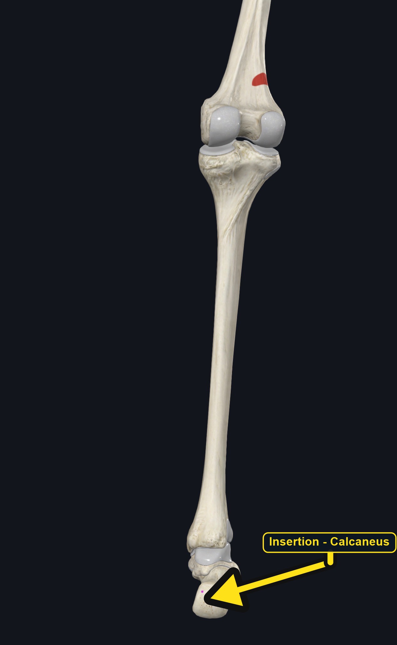
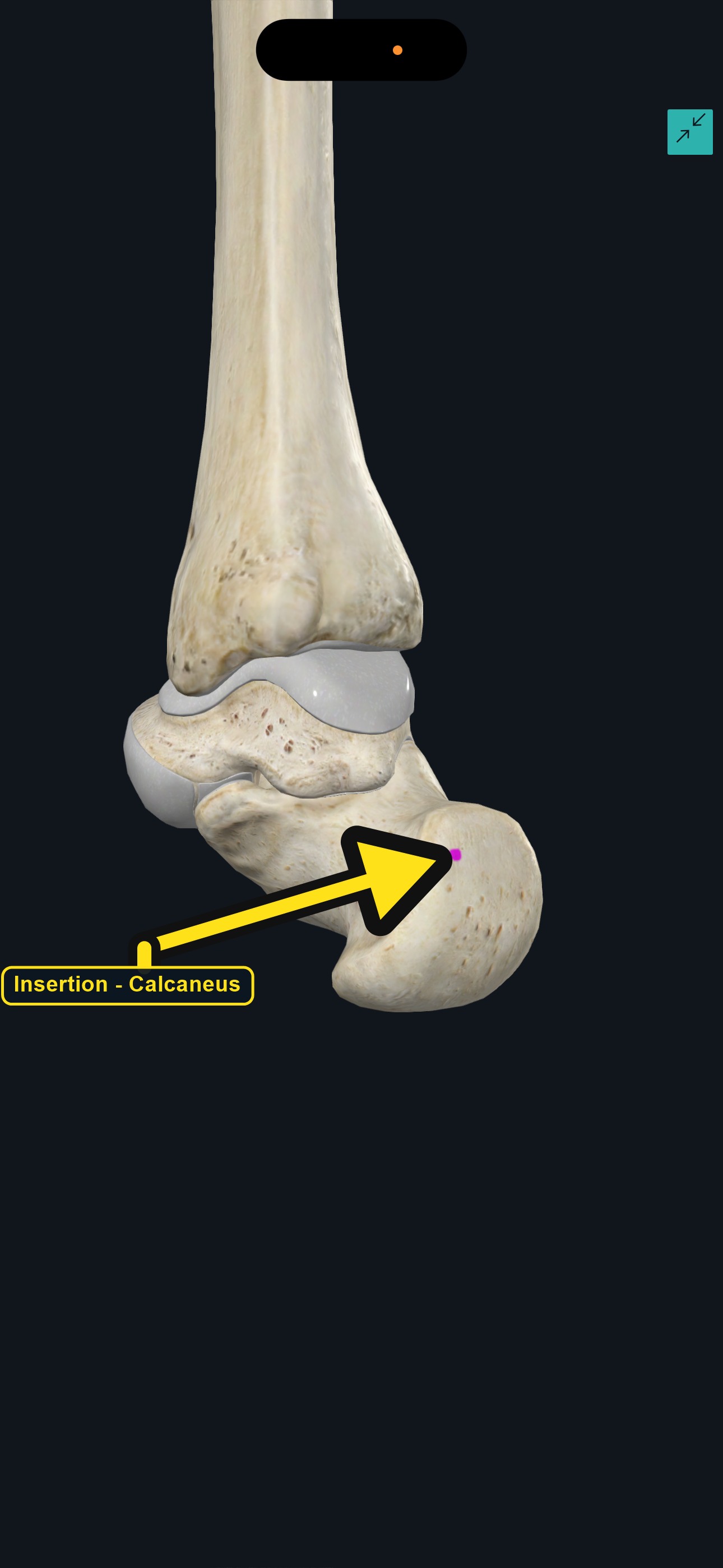
Insertion
| Insertion | The posterior surface of the calcaneus, medial to the Achilles tendon, or blending into the Achilles tendon (or the flexor retinaculum) in some people. |
|---|
Section 04 of 20
Actions
No dedicated action video is currently available. This movement is explained below and video will be added when verified source media becomes available.
| Primary action | Weak plantar flexion of the ankle. |
|---|---|
| Secondary actions | Weak knee flexion; a possible tensioning role at the back of the knee and heel. |
| Movement at the joint | Ankle joint and knee joint. |
| Heads / fiber groups | Single small belly with a long tendon. |
| Functional examples | Walking; running; rising onto the toes; push-off when jumping. |
Plantar flexion of the ankle
Weakly points the foot downward, working with gastrocnemius and soleus.
Knee flexion
Weakly assists bending the knee, because the belly crosses behind the knee joint.
Sensory and tension role
The long tendon may help tension the back of the knee and heel tissues; its exact role is debated.
Section 05 of 20
Nerve Innervation
| Nerve | Tibial nerve, roots S1–S2. |
|---|
The tibial nerve and popliteal vessels lie close to the belly in the back of the knee. Calf swelling, warmth, or sudden pain may reflect a blood clot or other serious problem, not only a muscle strain.
Section 06 of 20
Blood Supply
| Blood | Sural arteries and branches of the popliteal artery. |
|---|
Section 07 of 20
Pictures and Visual Anatomy
Section 08 of 20
Functional Movement
Plantaris assists during walking, running, and rising onto the toes, when the calf muscles push the body forward. Because it is small and sometimes absent, its functional contribution is minor.
Section 09 of 20
Synergists and Antagonists
Synergists: gastrocnemius and soleus (plantar flexion), and the hamstrings and popliteus (knee flexion). Antagonists: the tibialis anterior and other ankle dorsiflexors (dorsiflexion), and the quadriceps (knee extension).
Section 10 of 20
Palpation and Location
Client position and technique
The muscle cannot be felt separately. Position the client prone with the leg supported and the foot resting relaxed. Locate the lateral head of gastrocnemius behind the knee and the border between it and the soleus, then use gentle, broad pressure to feel the area where the belly lies. Ask for gentle plantar flexion to feel the overlying gastrocnemius contract.
Protect nearby structures
The popliteal fossa contains the popliteal artery and vein, the tibial and common fibular nerves, and lymph nodes. Avoid deep or sustained pressure behind the knee, and do not massage a swollen, warm, or tender calf. Stop if the client reports tingling, numbness, or sharp pain.
Section 11 of 20
Massage Therapy Relevance
Plantaris is discussed in calf pain and Achilles-region problems in runners and jumpers. Its rupture is sometimes mistaken for a blood clot in the calf, so careful screening matters, and many other conditions cause calf pain.
Section 12 of 20
Dysfunction: What Happens When Movement Goes Wrong
Changes in how a muscle works can change how a joint moves. The points below describe possibilities that may be associated with altered muscle function; they are not diagnoses, and one muscle is never assumed to be the sole cause of a problem.
When the muscle is weak
Weakness is difficult to detect because the muscle is small and may be absent; plantar flexion strength is mainly carried by gastrocnemius and soleus.
When it is tight or shortened
Tightness may be associated with posterior knee discomfort and limited ankle dorsiflexion with the knee extended.
When it is overused
Running, jumping, and repeated push-off may overload the calf muscles and may contribute to calf and Achilles-region pain.
When it is strained
A strain may cause a sudden sharp calf pain during push-off; gastrocnemius or soleus injury and a blood clot can look similar.
When it is inhibited or others compensate
Reduced activation may be associated with more work by gastrocnemius and soleus.
When it does not coordinate well with synergists and antagonists
Poor coordination of the calf muscles may alter push-off and may be associated with calf or Achilles symptoms.
Section 13 of 20
Conditions, Pathologies, and Pain Relationships
Plantaris strain or rupture
Sudden pain in the calf, often described as a snap during a push-off or a sudden stop, may involve plantaris (sometimes called tennis leg). Gastrocnemius and soleus injuries look similar, and assessment is needed.
Achilles tendinopathy
Pain and thickening of the mid Achilles tendon may sometimes involve the plantaris tendon lying next to it, but many other structures are involved.
Calf tightness and cramps
Tight calf muscles may be associated with posterior knee and heel discomfort.
Differential considerations
Deep vein thrombosis, Baker cyst rupture, gastrocnemius or soleus strain, popliteal artery entrapment, and lumbar or sciatic referral can all cause calf pain.
Section 14 of 20
Example Clinical Relationship
A recreational tennis player reports a sudden sharp pain in the back of the calf when pushing off, followed by swelling and bruising. Plantaris, gastrocnemius, or soleus may be involved, and the pattern may be associated with a calf strain, but a blood clot can look similar. Because of that risk, a swollen, warm, or painful calf needs medical assessment before any massage.
Section 15 of 20
Massage and Treatment Approaches
For healthy, non-acute tissue, position the client prone with the ankle supported. Begin with light effleurage from the ankle toward the knee, then use gentle kneading of the gastrocnemius and soleus, keeping pressure light behind the knee and never pressing into the popliteal fossa.
| Movement | Use gentle ankle plantar flexion and dorsiflexion and comfortable calf stretching; do not force range. |
|---|---|
| Regional work | Consider gastrocnemius, soleus, the Achilles tendon area, and the hamstrings when assessment supports it. |
| Avoid | Deep pressure behind the knee, over a swollen, warm, or bruised calf, or over an acute injury. |
Section 16 of 20
Precautions and Contraindications
Precautions and contraindications
- Acute calf injury, swelling, bruising, or suspected blood clot: do not massage, and refer.
- Recent knee, calf, or Achilles injury or surgery.
- Use caution with varicose veins, anticoagulant use, or easy bruising.
- Stop if pressure causes sharp pain, tingling, or numbness.
Section 17 of 20
Red Flags and When to Refer
Red flags and referral
- A swollen, warm, red, or painful calf, especially on one side, or sudden shortness of breath or chest pain (possible blood clot).
- A sudden snap with severe calf pain, swelling, and bruising, or inability to rise onto the toes.
- A cold, pale, or numb foot, or loss of pulse.
- Calf pain with fever, or after a long flight or period of immobility.
Section 18 of 20
Study Summary
Attachments
Origin: lateral supracondylar line of the femur and oblique popliteal ligament. Insertion: posterior calcaneus, medial to the Achilles tendon.
Actions
Weak plantar flexion of the ankle and weak knee flexion.
Supply
Tibial nerve (S1–S2); sural and popliteal artery branches.
Section 19 of 20
Instructor Narration
Open narration transcript
Plantaris is a small calf muscle with a very long, thin tendon. It is absent in a small percentage of people.
It arises from the lateral supracondylar line of the femur and the oblique popliteal ligament and inserts on the posterior calcaneus, medial to the Achilles tendon.
It weakly plantar flexes the ankle and weakly flexes the knee. Gastrocnemius and soleus help it, and the ankle dorsiflexors and the quadriceps oppose it.
The tibial nerve supplies it, from S1 to S2, and the sural and popliteal artery branches provide its blood.
It cannot be felt on its own. Work gently on the calf, avoid the popliteal fossa, and never massage a swollen, warm, or painful calf.
A plantaris or gastrocnemius strain, Achilles tendinopathy, and calf problems can overlap, and a blood clot can look like a strain. Refer a swollen warm calf, a sudden snap with severe pain and bruising, a cold or numb foot, or chest pain with shortness of breath.
Remember: lateral supracondylar line of the femur to the calcaneus; weak plantar flexion and knee flexion; tibial nerve.
Section 20 of 20