Section 01 of 20
Introduction
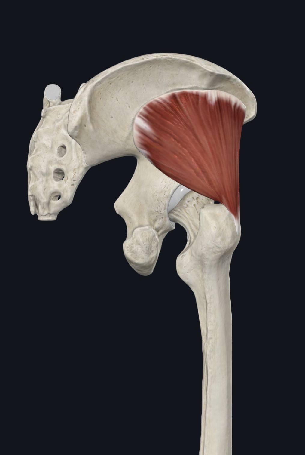
Gluteus minimus is the smallest and deepest of the three gluteal muscles. It lies directly beneath gluteus medius on the outer surface of the ilium, and its fibers converge like a fan toward the greater trochanter of the femur.
Together with gluteus medius and tensor fasciae latae it forms the lateral hip stabilizer group, and its tendon lies close to the hip joint capsule and the trochanteric bursa.
Section 02 of 20
Origin
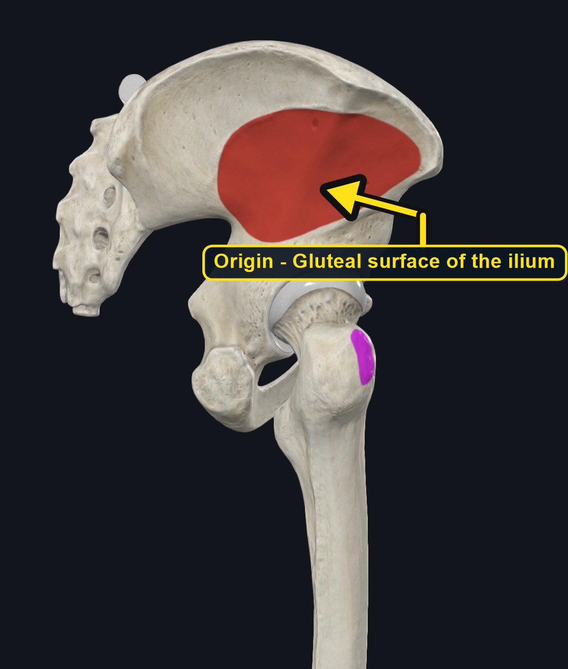
| Origin | External (gluteal) surface of the ilium between the anterior and inferior gluteal lines, and the margin of the greater sciatic notch. |
|---|
Section 03 of 20
Insertion
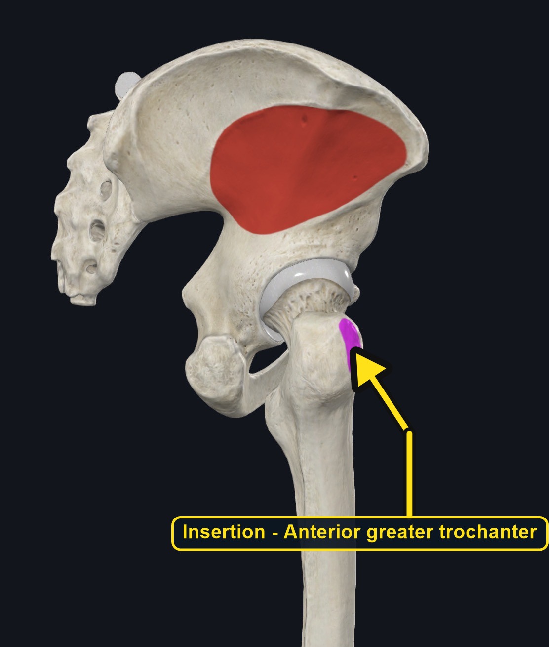
| Insertion | Anterior surface of the greater trochanter of the femur, with fibers blending into the hip joint capsule. |
|---|
Section 04 of 20
Actions
No dedicated action video is currently available. This movement is explained below and video will be added when verified source media becomes available.
| Primary action | Hip abduction. |
|---|---|
| Secondary actions | Medial (internal) rotation and weak flexion by the anterior fibers; stabilizes the pelvis in single-leg stance. |
| Movement at the joint | Hip joint. |
| Heads / fiber groups | Anterior and posterior fiber groups; the anterior fibers rotate the hip inward. |
| Functional examples | Walking and running (keeping the pelvis level); stepping sideways; standing on one leg; climbing stairs. |
Hip abduction
Draws the thigh away from the midline, especially with gluteus medius.
Medial rotation
The anterior fibers rotate the thigh inward and assist hip flexion.
Pelvic and joint stability
Holds the pelvis level in single-leg stance and helps keep the femoral head seated in the socket.
Section 05 of 20
Nerve Innervation
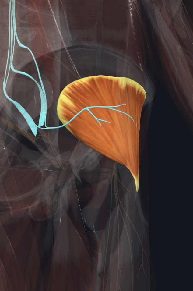
| Nerve | Superior gluteal nerve, roots L4–S1. |
|---|
The superior gluteal nerve and vessels run between gluteus medius and gluteus minimus. Weakness of hip abduction with a pelvic drop on the opposite side may reflect nerve or joint problems, not only muscle tightness.
Section 06 of 20
Blood Supply
| Blood | Deep branch of the superior gluteal artery, with contributions from the inferior gluteal artery. |
|---|
Section 07 of 20
Pictures and Visual Anatomy
Section 08 of 20
Functional Movement
Gluteus minimus works with every step of walking and running, keeping the pelvis from dropping on the swinging side, and stabilizes the hip when you stand on one leg, climb stairs, or step sideways.
Section 09 of 20
Synergists and Antagonists
Synergists: gluteus medius and tensor fasciae latae (abduction and medial rotation). Antagonists: the hip adductors (adductor longus, brevis, and magnus, gracilis, and pectineus) and the lateral rotators.
Section 10 of 20
Palpation and Location
Client position and technique
Position the client side-lying with the upper leg supported and slightly flexed. The muscle lies under gluteus medius and cannot be felt separately, but firm, broad pressure above the greater trochanter and below the iliac crest reaches the gluteal region where it lies. Ask for gentle hip abduction to feel the overlying muscles contract.
Protect nearby structures
Use slow, broad pressure and stay off the greater trochanter itself, where the bursae and tendons are often tender. Avoid sustained pressure that sends pain or tingling down the leg, and keep away from the sciatic nerve path in the lower buttock.
Section 11 of 20
Massage Therapy Relevance
Lateral hip pain is common in runners, in people who stand on one leg or sleep on one side, and in older adults. Gluteus minimus and medius are frequent contributors, but hip joint, spine, and bursa problems can look the same.
Section 12 of 20
Dysfunction: What Happens When Movement Goes Wrong
Changes in how a muscle works can change how a joint moves. The points below describe possibilities that may be associated with altered muscle function; they are not diagnoses, and one muscle is never assumed to be the sole cause of a problem.
When the muscle is weak
Weakness may cause the pelvis to drop on the opposite side in single-leg stance and may be associated with a side-to-side gait and greater load on the low back and knee.
When it is tight or shortened
Tightness may be associated with lateral hip ache and limited hip adduction and internal rotation, and may add tension near the greater trochanter.
When it is overused
Running, long standing, and repeated single-leg loading may overload the muscle and its tendon and may contribute to lateral hip pain.
When it is strained
A strain or tendon irritation may cause outer-hip pain with side-lying or single-leg stance; hip joint disease and lumbar referral can look similar.
When it is inhibited or others compensate
Reduced activation may be associated with inward knee collapse during squats or landing and more work by tensor fasciae latae and the low back.
When it does not coordinate well with synergists and antagonists
Poor coordination with gluteus medius, gluteus maximus, and the adductors may alter pelvic and knee control and may be associated with lateral hip or knee symptoms.
Section 13 of 20
Conditions, Pathologies, and Pain Relationships
Gluteal tendinopathy and greater trochanteric pain syndrome
Pain over the outer hip, worse when lying on that side or standing on one leg, may involve the gluteal tendons and bursae. Deep pressure on the trochanter is not appropriate.
Weakness and pelvic drop
Weak abductors may allow the pelvis to drop on the opposite side when walking (a Trendelenburg pattern) and may contribute to lateral hip or low back symptoms.
Trigger point referral
Deep pain patterns that spread down the outer thigh and leg may involve the gluteal muscles, but they can also come from the lumbar spine (especially L5).
Differential considerations
Hip osteoarthritis, femoroacetabular impingement, lumbar radiculopathy, sacroiliac problems, and fractures can all cause lateral hip and buttock pain.
Section 14 of 20
Example Clinical Relationship
A runner reports outer-hip pain that is worse when lying on that side at night. The deep gluteal tendons and bursa may be involved, and the pattern may be associated with gluteal tendinopathy, but the hip joint and the lower back can cause similar pain. Gentle broad work around the trochanter may support comfort, while sudden severe pain after a fall or pain with fever needs referral.
Section 15 of 20
Massage and Treatment Approaches
For healthy, non-acute tissue, position the client side-lying with pillows between the knees. Begin with broad effleurage over the gluteal region and progress to slow compression and kneading of the upper buttock and iliac crest border, staying within comfortable pressure.
| Movement | Use gentle active hip abduction and comfortable range-of-motion work; do not force rotation. |
|---|---|
| Regional work | Consider gluteus medius and maximus, tensor fasciae latae, piriformis, and quadratus lumborum when assessment supports it. |
| Avoid | Deep pressure on the greater trochanter, an acutely painful tendon, or any point that reproduces leg symptoms. |
Section 16 of 20
Precautions and Contraindications
Precautions and contraindications
- Acute hip injury, recent hip surgery or replacement, or suspected fracture.
- Use caution with severe osteoporosis, anticoagulant use, or easy bruising.
- Avoid open wounds, infection, and unexplained swelling.
- Stop if pressure produces sharp pain or leg tingling.
Section 17 of 20
Red Flags and When to Refer
Red flags and referral
- Sudden hip pain after a fall, or inability to bear weight.
- Fever, night pain, or unexplained weight loss with hip or buttock pain.
- Progressive leg weakness, foot drop, or numbness in the saddle area.
- Bladder or bowel changes with back or leg symptoms (possible cauda equina syndrome).
Section 18 of 20
Study Summary
Attachments
Origin: gluteal surface of the ilium. Insertion: anterior greater trochanter.
Actions
Hip abduction and medial rotation; stabilizes the pelvis.
Supply
Superior gluteal nerve (L4–S1); superior gluteal artery.
Section 19 of 20
Instructor Narration
Open narration transcript
Gluteus minimus is the smallest and deepest of the gluteal muscles. It lies beneath gluteus medius and fans out from the outer surface of the ilium toward the greater trochanter.
It arises from the gluteal surface of the ilium between the anterior and inferior gluteal lines and inserts on the anterior surface of the greater trochanter, blending into the hip joint capsule.
It abducts the thigh, the anterior fibers medially rotate the hip, and the muscle helps keep the pelvis level when you stand on one leg. Gluteus medius and tensor fasciae latae work with it, and the adductors oppose it.
The superior gluteal nerve supplies it, from L4 to S1, and the superior gluteal artery provides its blood.
It cannot be felt on its own, so position the client side-lying and use broad, slow pressure above the trochanter, staying off the trochanter itself and away from anything that sends pain down the leg.
Gluteal tendinopathy, trochanteric pain, weakness with a pelvic drop, hip arthritis, and lumbar nerve problems can all overlap. Refer sudden hip pain after a fall, fever or night pain, progressive leg weakness, or bladder or bowel changes.
Remember: gluteal surface of the ilium to the anterior greater trochanter, hip abduction and medial rotation, superior gluteal nerve.
Section 20 of 20