Section 01 of 20
Introduction
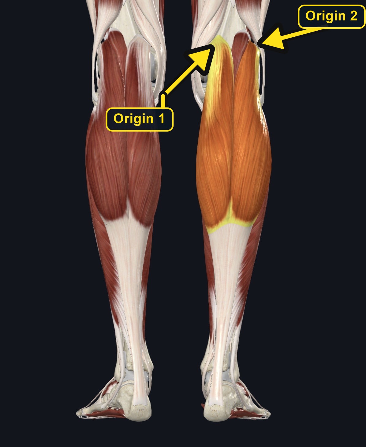
Gastrocnemius forms the visible bulk of the calf. Its medial and lateral heads arise from the femur above the knee, so the muscle crosses both the knee and the ankle.
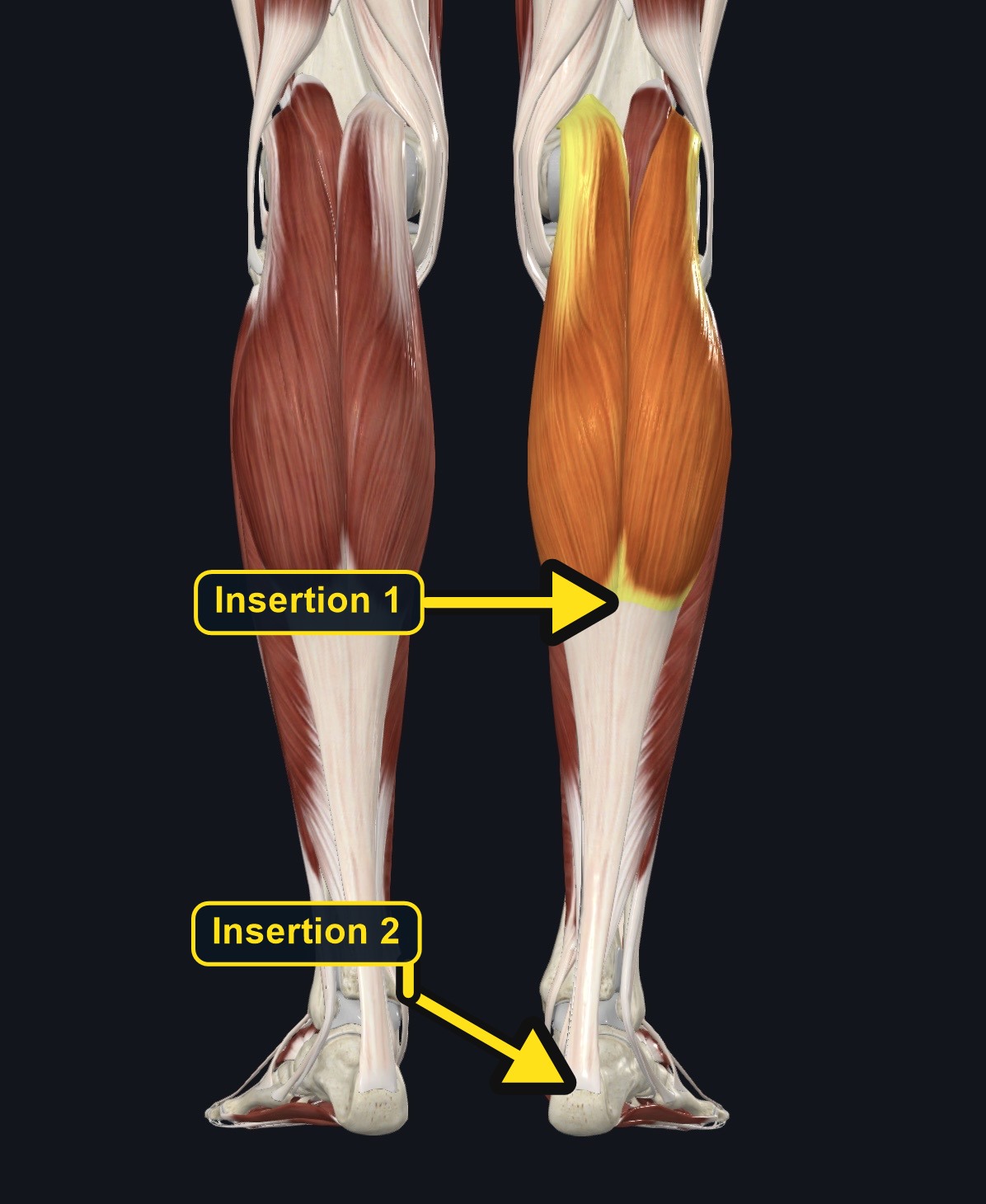
The two heads unite and join the soleus tendon to form the Achilles (calcaneal) tendon, which inserts on the back of the heel bone.
Section 02 of 20
Origin
| Origin | Medial head: popliteal surface of the femur just above the medial condyle. Lateral head: lateral surface of the lateral femoral condyle. |
|---|
Section 03 of 20
Insertion
| Insertion | Posterior surface of the calcaneus through the Achilles tendon, together with soleus. |
|---|
Section 04 of 20
Actions
| Primary action | Ankle plantarflexion (strongest with the knee straight). |
|---|---|
| Secondary actions | Knee flexion; assists propulsion and knee stability. |
| Movement at the joint | Ankle (talocrural) joint and knee joint. |
| Heads / fiber groups | Two heads: medial and lateral, both arising from the femoral condyles. |
| Functional examples | Rising onto the toes; walking and running push-off; jumping; climbing stairs. |
Ankle plantarflexion
Raises the heel, most strongly when the knee is extended.
Knee flexion
Assists knee flexion because the heads cross the knee.
Propulsion and stability
Provides push-off in walking, running, and jumping and stabilizes the knee and ankle.
Section 05 of 20
Nerve Innervation
| Nerve | Tibial nerve, roots S1–S2. |
|---|
The tibial nerve and popliteal vessels lie deep to the two heads in the popliteal fossa. Calf symptoms with numbness, weakness, or swelling may reflect nerve or vascular problems, not muscle tightness.
Section 06 of 20
Blood Supply
| Blood | Sural arteries from the popliteal artery, with contributions from the posterior tibial artery. |
|---|
Section 07 of 20
Pictures and Visual Anatomy
The pictures for this muscle are shown with the Origin, Insertion, Nerve Innervation, and other sections where they teach best.
Section 08 of 20
Functional Movement
Gastrocnemius is central to walking, running, jumping, stair climbing, and rising onto the toes. It works best with the knee extended, while soleus is more active with the knee bent.
Section 09 of 20
Synergists and Antagonists
Synergists: soleus, plantaris, tibialis posterior, and the fibularis muscles in plantarflexion; the hamstrings in knee flexion. Antagonists: tibialis anterior and the toe extensors in ankle dorsiflexion; the quadriceps in knee extension.
Section 10 of 20
Palpation and Location
Client position and technique
Position the client prone with the ankle supported and the knee slightly flexed. Ask them to point the foot down gently against light resistance, or to rise onto their toes in standing, to see and feel the two bellies. Trace each head up toward the femoral condyles, and follow the muscle down to the Achilles tendon.
Protect nearby structures
Avoid deep or sustained pressure in the popliteal fossa behind the knee, where the tibial nerve and popliteal vessels are close to the surface. Do not massage a calf that is swollen, hot, red, or painful without a known cause, and avoid strong pressure on the Achilles tendon.
Section 11 of 20
Massage Therapy Relevance
Calf tightness and soreness are very common in runners, people who stand for long periods, and people who wear heels. Because calf pain can also signal a clot or other serious problem, screening is essential.
Section 12 of 20
Dysfunction: What Happens When Movement Goes Wrong
Changes in how a muscle works can change how a joint moves. The points below describe possibilities that may be associated with altered muscle function; they are not diagnoses, and one muscle is never assumed to be the sole cause of a problem.
When the muscle is weak
Weakness may reduce push-off strength and may be associated with a shorter stride and more load on the soleus and the Achilles tendon.
When it is tight or shortened
Tightness may be associated with limited ankle dorsiflexion and may contribute to heel lift, early heel rise, or overpronation.
When it is overused
Running, long standing, and high heels may overload the muscle and may contribute to calf ache or Achilles tendon irritation.
When it is strained
A strain may cause sudden sharp pain in the calf during push-off, often in the medial head, with swelling or bruising; acute injury needs assessment.
When it is inhibited or others compensate
Reduced activation may be associated with weaker push-off and more work by the hamstrings and hip.
When it does not coordinate well with synergists and antagonists
Poor coordination with the soleus, tibialis anterior, and hamstrings may alter gait and knee control and may increase load on the Achilles tendon.
Section 13 of 20
Conditions, Pathologies, and Pain Relationships
Calf strain (tennis leg)
A sudden sharp pain in the medial calf during push-off, often at the medial head's musculotendinous junction. Acute pain, bruising, or swelling needs assessment rather than deep work.
Achilles tendinopathy
Pain and stiffness in the tendon or heel-cord area, especially in the morning or after activity. Avoid aggressive pressure on the tendon.
Cramps and overuse
Muscle cramps and aching can follow heavy exercise, dehydration, or long standing and often settle with rest and gentle work.
Differential considerations
Deep vein thrombosis, Baker cyst rupture, compartment syndrome, S1 radiculopathy, peripheral arterial disease, and Achilles tendon rupture can all present as calf pain.
Section 14 of 20
Example Clinical Relationship
A recreational runner reports sudden pain in the calf while pushing off, with swelling. The gastrocnemius may be strained (sometimes called tennis leg), but calf pain can also come from a blood clot, a ruptured Baker cyst, or compartment syndrome. Unilateral swelling, warmth, redness, chest pain, or shortness of breath needs urgent referral.
Section 15 of 20
Massage and Treatment Approaches
For healthy, non-acute tissue, position the client prone with the ankle supported. Begin with broad effleurage and kneading over both heads, then progress to gentle compression and comfortable longitudinal strokes toward the Achilles tendon, staying out of the popliteal fossa.
| Movement | Use gentle active ankle pumping and comfortable passive dorsiflexion within pain-free range. |
|---|---|
| Regional work | Consider soleus, the hamstrings, the plantar fascia, and the foot when assessment supports it. |
| Avoid | Deep pressure on an acute strain, the popliteal fossa, the Achilles tendon, or a calf with unexplained swelling. |
Section 16 of 20
Precautions and Contraindications
Precautions and contraindications
- Acute strain, hematoma, recent surgery, or a recent long trip or immobilization with calf pain.
- Use caution with varicose veins, anticoagulant use, diabetes, or reduced sensation.
- Avoid open wounds, infection, and marked swelling.
- Do not massage a calf with unexplained unilateral swelling or warmth.
Section 17 of 20
Red Flags and When to Refer
Red flags and referral
- Unilateral calf swelling, warmth, redness, or pain, especially after immobility, surgery, or a long trip (possible deep vein thrombosis).
- Sudden shortness of breath, chest pain, or coughing blood.
- A sudden snap at the back of the ankle with inability to push off (possible Achilles tendon rupture).
- A tense, severely painful calf after injury, or cold, pale, or numb foot.
Section 18 of 20
Study Summary
Attachments
Origin: medial and lateral femoral condyles. Insertion: calcaneus via the Achilles tendon.
Actions
Ankle plantarflexion and knee flexion.
Supply
Tibial nerve (S1-S2); sural arteries.
Section 19 of 20
Instructor Narration
Open narration transcript
Gastrocnemius is the two-headed superficial muscle of the calf. Its medial and lateral heads arise from the femur just above the knee, so it crosses both the knee and the ankle.
The heads join the soleus tendon to form the Achilles tendon, which inserts on the back of the calcaneus.
Gastrocnemius plantarflexes the ankle, most strongly with the knee extended, and it assists knee flexion. Soleus and plantaris work with it, and tibialis anterior opposes ankle plantarflexion.
The tibial nerve, from S1 to S2, supplies it, and the sural arteries provide blood.
To palpate, place the client prone with the ankle supported and ask for a gentle downward foot press. Trace each head up to the knee and down to the Achilles tendon. Avoid the popliteal fossa, where the tibial nerve and popliteal vessels lie close to the surface.
Calf strain, Achilles tendinopathy, cramps, deep vein thrombosis, Baker cyst rupture, compartment syndrome, and nerve root problems can all cause calf pain. Refer unilateral calf swelling or warmth, sudden shortness of breath or chest pain, a snap at the back of the ankle, or a tense, painful calf after injury.
Remember: femoral condyles to the calcaneus by the Achilles tendon, plantarflexion and knee flexion, and the tibial nerve.
Section 20 of 20