Section 01 of 20
Introduction
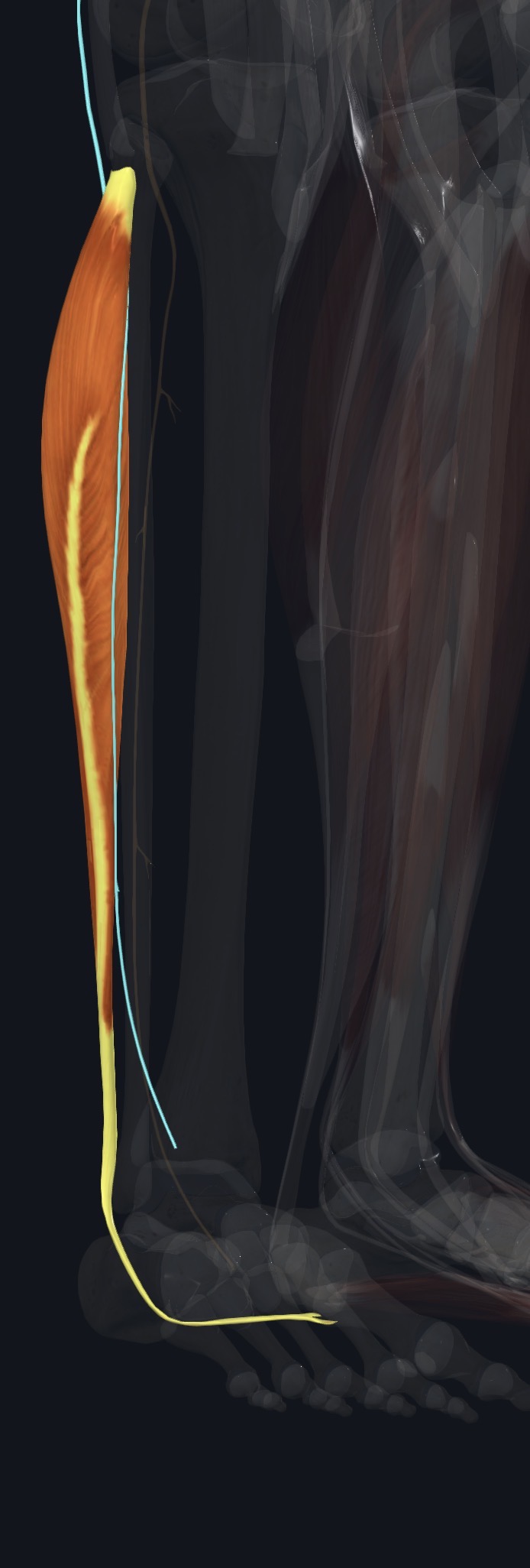
Fibularis longus—also called peroneus longus—is the more superficial and proximal of the two principal muscles in the lateral compartment of the leg. Its long tendon passes behind the lateral malleolus, crosses the sole through the cuboid groove, and reaches the medial side of the forefoot.
Section 02 of 20
Origin
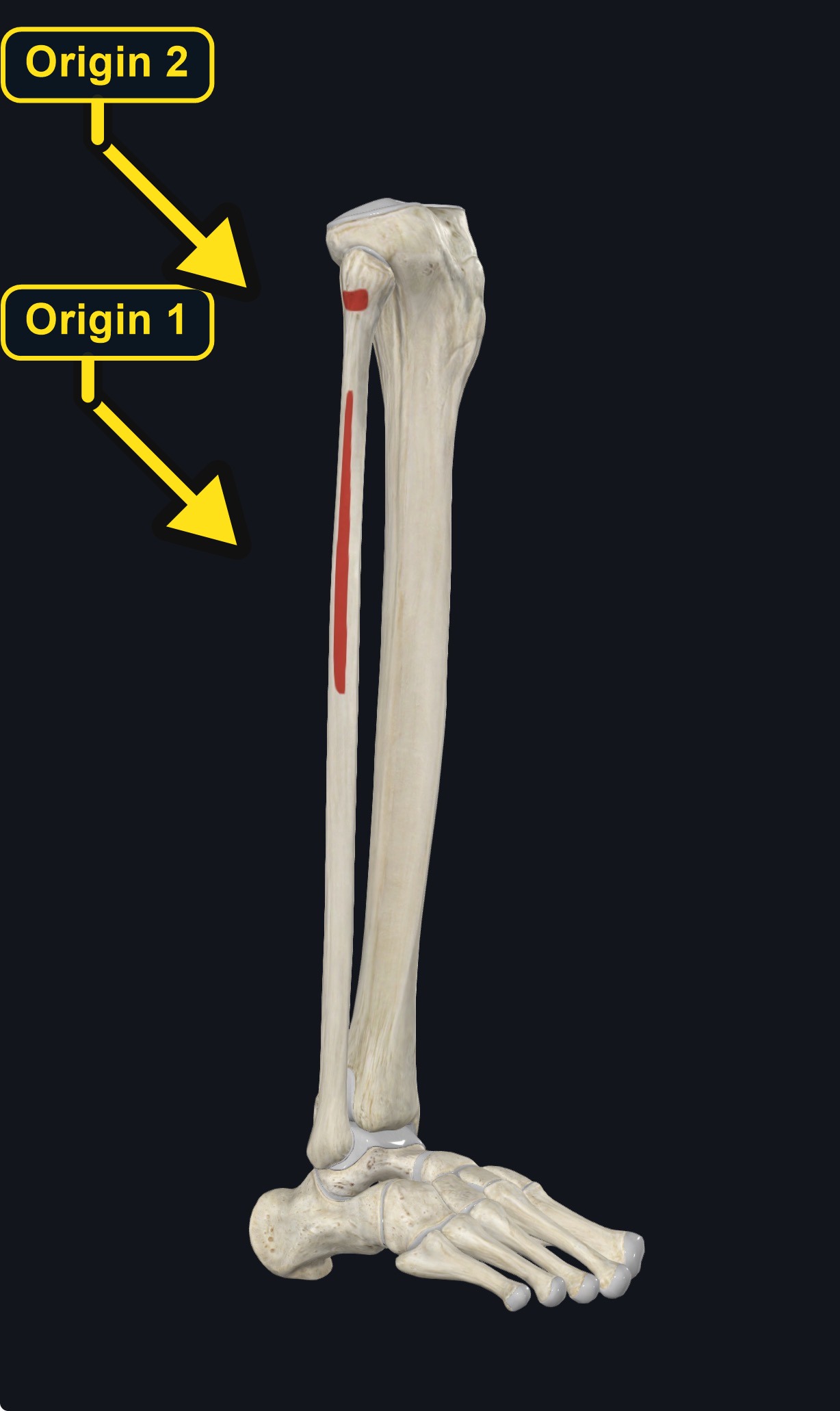
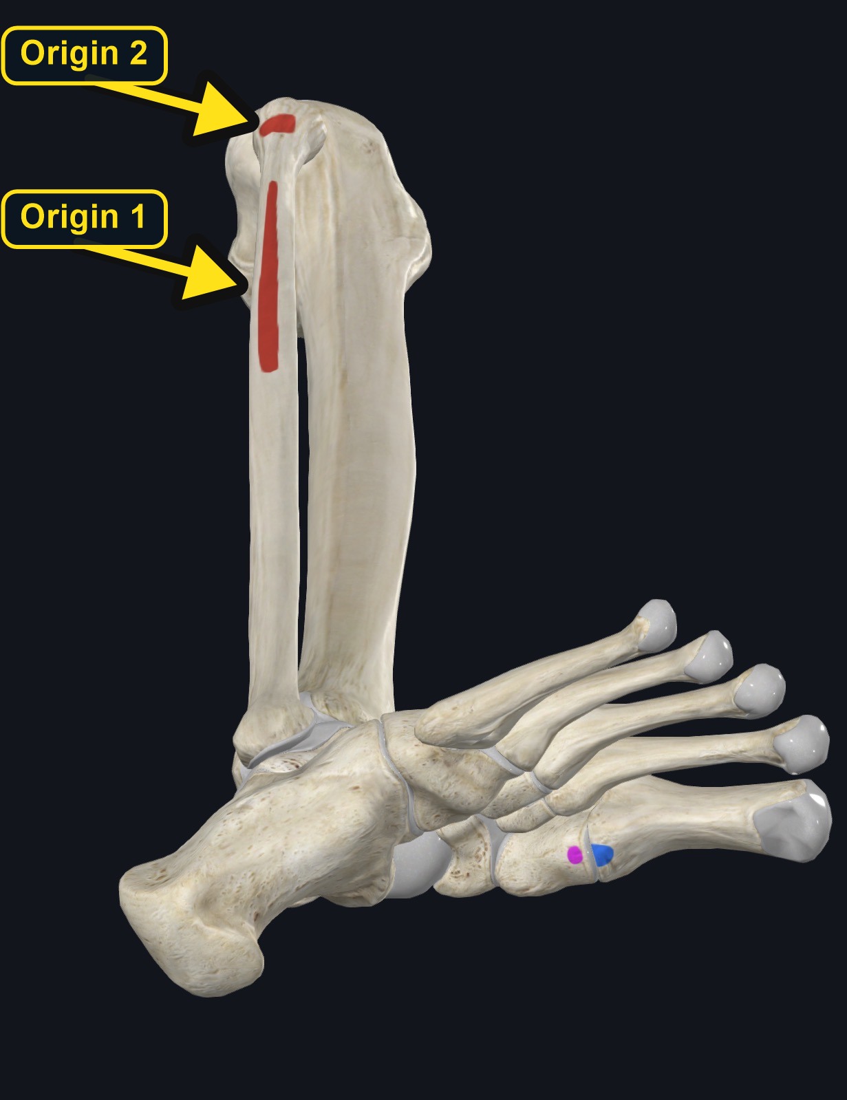
| Origin | Head and proximal two-thirds of the lateral fibula, with attachments to the lateral tibial condyle, deep fascia, and intermuscular septa. |
|---|
Section 03 of 20
Insertion
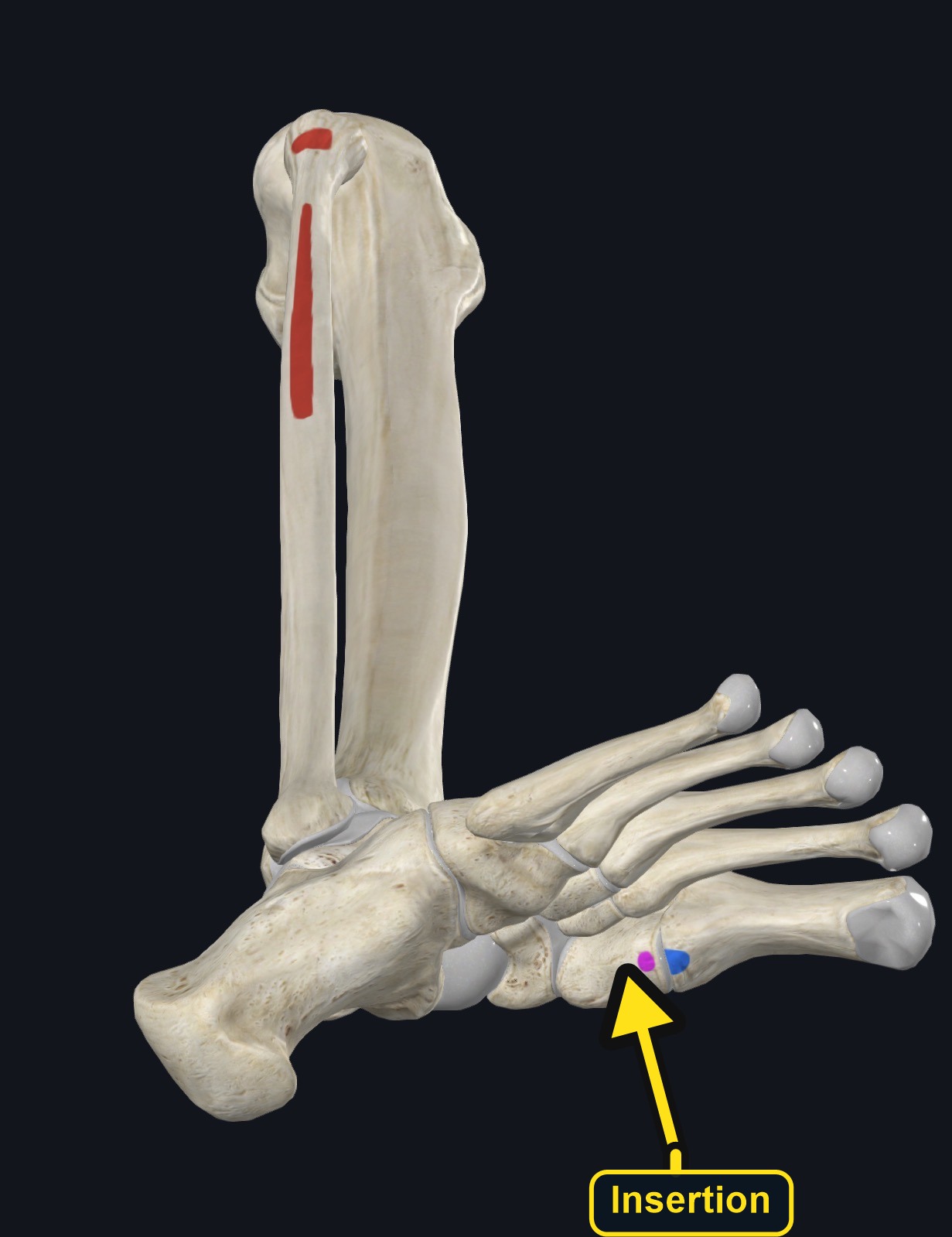
| Insertion | Plantar surface of the medial cuneiform and base of the first metatarsal after the tendon crosses the sole. |
|---|
The common fibular nerve curves around the fibular neck near the proximal attachment and requires particular caution.
Section 04 of 20
Actions
| Primary action | Eversion of the foot. |
|---|---|
| Secondary actions | Assists ankle plantarflexion; stabilizes the first ray and supports the transverse and lateral longitudinal arches. |
| Movement at the joint | Subtalar and transverse tarsal joints (eversion); ankle (weak plantarflexion). |
| Heads / fiber groups | Single muscle belly with a long tendon that crosses the sole. |
| Functional examples | Adapting the foot to uneven ground; controlling inversion during walking and running; single-leg balance; push-off through the big toe. |
- Everts the foot at the subtalar and transverse-tarsal joints.
- Assists ankle plantarflexion.
- Helps stabilize the first ray and supports the transverse and lateral longitudinal arches during weight bearing.
Section 05 of 20
Nerve Innervation
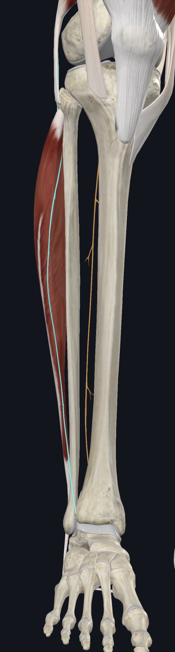
| Nerve | Superficial fibular (peroneal) nerve. |
|---|---|
| Roots | Primarily L5–S1; broader L5–S2 contribution is described in some references. |
| Key relationships | The common fibular nerve is vulnerable at the fibular neck; the superficial fibular nerve travels within the lateral compartment before becoming cutaneous distally. |
Section 06 of 20
Blood Supply
| Blood supply | Branches of the fibular artery, with contributions from the anterior tibial circulation. |
|---|
Section 07 of 20
Pictures and Visual Anatomy
Section 08 of 20
Functional Movement
Functionally, fibularis longus helps adapt the foot to the ground, controls inversion stresses, and stabilizes the foot during walking, running, and single-leg balance.
Section 09 of 20
Synergists and Antagonists
Synergists: fibularis brevis for eversion and the calf plantarflexors for plantarflexion. Antagonists: tibialis anterior and tibialis posterior oppose eversion; ankle dorsiflexors oppose plantarflexion.
Section 10 of 20
Palpation and Location
With the client side-lying, supine, or seated and the knee supported, locate the lateral fibular shaft. Ask for gentle eversion with slight plantarflexion; the proximal muscle belly firms below the fibular head. Follow the tendon distally behind the lateral malleolus, distinguishing it from fibularis brevis. Do not press directly over the fibular neck or aggressively compress the lateral compartment.
Section 11 of 20
Massage Therapy Relevance
Fibularis longus may be loaded by running, uneven terrain, repeated ankle stabilization, footwear changes, or recovery from inversion sprain. Lateral-leg or ankle pain is not automatically muscular; tendon injury, joint or ligament injury, nerve dysfunction, fracture, vascular problems, and exertional compartment disorders can overlap.
Section 12 of 20
Dysfunction: What Happens When Movement Goes Wrong
Changes in how a muscle works can change how a joint moves. The points below describe possibilities that may be associated with altered muscle function; they are not diagnoses, and one muscle is never assumed to be the sole cause of a problem.
When the muscle is weak
Weakness may reduce control of inversion, may be associated with ankle instability, and may increase the load on the ankle ligaments.
When it is tight or shortened
Tightness may be associated with a lateral leg ache and may restrict foot inversion and dorsiflexion.
When it is overused
Running on uneven ground or repeated ankle stabilization may overload the muscle and may contribute to lateral leg or ankle pain.
When it is strained
A strain or tendon injury may cause pain behind the lateral malleolus with resisted eversion; ankle pain also has ligament, joint, and bone causes.
When it is inhibited or others compensate
Reduced activation, for example after an ankle sprain, may be associated with poorer balance and a tendency to roll the ankle.
When it does not coordinate well with synergists and antagonists
Poor coordination with fibularis brevis, tibialis posterior, and the calf may alter foot stability and arch support.
Section 13 of 20
Conditions, Pathologies, and Pain Relationships
Fibularis tendinopathy or strain
May produce lateral-leg or posterolateral-ankle pain aggravated by resisted eversion or prolonged loading. Massage may support comfort in a non-acute presentation but does not repair a tear.
Ankle sprain and instability
The evertors help resist inversion, but persistent instability, swelling, or inability to bear weight needs assessment rather than repeated provocation.
Tendon subluxation or tear
Painful snapping behind the lateral malleolus, acute weakness, or traumatic onset warrants medical evaluation.
Nerve or compartment involvement
Foot drop, spreading numbness, progressive weakness, or severe tense lateral-leg pain can reflect nerve compromise or compartment syndrome.
Section 14 of 20
Example Clinical Relationship
A trail runner who has had an ankle sprain reports aching along the outer leg and pain behind the outer ankle bone. Fibularis longus may be overloaded and may be associated with tendon irritation or weakness after the sprain, but fractures, ligament injury, and nerve problems can look similar. Sudden swelling, inability to bear weight, or numbness needs referral.
Section 15 of 20
Massage and Treatment Approaches
Assess comfort, recent injury, swelling, and neurologic symptoms before treatment. Use broad, tolerable work along the lateral leg and avoid direct sustained pressure at the fibular neck, aggressive compression of an irritable tendon behind the lateral malleolus, or attempts to reproduce nerve symptoms. Massage therapists do not diagnose tendon tears, instability, neuropathy, or compartment syndrome.
- Broad warming strokes to the lateral lower leg within comfort.
- Gentle longitudinal work along the accessible muscle belly.
- Comfortable inversion and dorsiflexion to lengthen the muscle-tendon unit without forcing range.
- Side-lying, supine, or seated positioning with the knee and ankle supported.
- Reassess symptoms after changing ankle position or pressure.
Section 16 of 20
Precautions and Contraindications
Avoid local massage with acute trauma, suspected fracture or tendon rupture, infection, open wounds, acute unexplained swelling, DVT concern, recent surgery without clearance, or severe neurologic symptoms. Use special caution near the fibular head and with anticoagulation, fragile skin, or marked sensory loss.
Section 17 of 20
Red Flags and When to Refer
Refer urgently for severe escalating pain with a tense compartment, rapidly increasing swelling, pale or cool foot, loss of pulses, major trauma, or sudden motor/sensory change. Prompt assessment is also appropriate for foot drop, progressive weakness or numbness, painful tendon snapping after injury, inability to bear weight, fever/redness, or persistent unexplained symptoms.
Section 18 of 20
Study Summary
Origin: fibular head and proximal lateral fibula. Insertion: plantar medial cuneiform and base of first metatarsal. Actions: eversion, assists plantarflexion, supports the arches. Innervation: superficial fibular nerve, primarily L5–S1. Clinical key: protect the common fibular nerve at the fibular neck and refer new weakness, foot drop, traumatic snapping, or compartment warning signs.
Section 19 of 20
Instructor Narration
Open complete narration
Section 20 of 20