Section 01 of 20
Introduction
Biceps femoris forms the lateral portion of the posterior thigh. Its long head crosses the hip and knee; its short head crosses only the knee. The heads unite distally and help form the superolateral border of the popliteal fossa.
The long head meets the usual hamstring definition because it begins at the ischial tuberosity and crosses two joints. The short head is functionally grouped with the hamstrings but begins on the femur.
Section 02 of 20
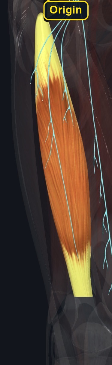
Origin
| Long-head origin | Ischial tuberosity, commonly through a proximal tendon shared with semitendinosus. |
|---|---|
| Short-head origin | Lateral lip of the linea aspera and lateral supracondylar line of the femur, with adjacent lateral intermuscular septum. |
Section 03 of 20
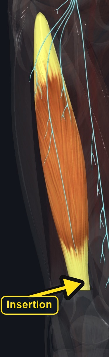
Insertion
| Insertion | Primarily the lateral side of the fibular head, with fascial and lateral tibial expansions described in detailed anatomy. |
|---|
Section 04 of 20
Actions
| Primary action | Knee flexion (both heads). |
|---|---|
| Secondary actions | Lateral rotation of the flexed leg; the long head also extends the hip and assists lateral rotation of the thigh. |
| Movement at the joint | Knee (flexion, lateral rotation) and hip (extension, long head only). |
| Heads / fiber groups | Long head (ischial tuberosity, crosses hip and knee) and short head (linea aspera and lateral supracondylar line, crosses the knee only), with different nerve supplies. |
| Functional examples | Walking and running (decelerating the swinging leg); rising from a chair; climbing; controlling forward bending of the trunk. |
Both heads
Flex the knee and laterally rotate the leg when the knee is flexed.
Long head
Extends the hip and assists lateral rotation of the thigh; eccentrically controls hip flexion and the forward-swinging leg.
Short head
Acts only at the knee because it does not cross the hip.
Section 05 of 20
Nerve Innervation
| Long head | Tibial division of the sciatic nerve, primarily L5–S2. |
|---|---|
| Short head | Common fibular division of the sciatic nerve, primarily L5–S2. |
The divided innervation is distinctive. Neurological symptoms may affect the heads differently and should not be assumed to reflect local muscle tightness.
Section 06 of 20
Blood Supply
| Blood | Perforating branches of the profunda femoris artery, with regional contributions from inferior gluteal and popliteal vessels. |
|---|
Section 07 of 20
Pictures and Visual Anatomy
The pictures for this muscle are shown with the Origin, Insertion, Nerve Innervation, and other sections where they teach best.
Section 08 of 20
Functional Movement
Walking, running, climbing, rising, decelerating the lower leg, and controlling forward trunk motion recruit biceps femoris.
Section 09 of 20
Synergists and Antagonists
Synergists: semitendinosus and semimembranosus in knee flexion; gluteus maximus and posterior adductor magnus in hip extension. Antagonists: quadriceps oppose knee flexion; iliopsoas and rectus femoris oppose hip extension.
Section 10 of 20
Palpation and Location
Client position
Position prone with the knee slightly flexed and supported, or side-lying. Ask for gentle knee flexion with slight lateral rotation against light resistance. Follow the lateral posterior-thigh belly toward its firm distal tendon at the fibular head.
Protect nearby structures
Stay out of the central popliteal fossa. The common fibular nerve winds around the fibular neck just distal to the insertion and is vulnerable to pressure. Avoid strong contact over the ischial tuberosity, popliteal space, and fibular neck.
Section 11 of 20
Massage Therapy Relevance
Biceps femoris is the lateral hamstring and a frequent site of soreness in runners, kickers, and people who sit for long periods. Its long head crosses the hip and knee, and its tendon reaches the fibular head where the common fibular nerve is close, so treatment involves careful pressure choices and screening for nerve symptoms.
Section 12 of 20
Dysfunction: What Happens When Movement Goes Wrong
Changes in how a muscle works can change how a joint moves. The points below describe possibilities that may be associated with altered muscle function; they are not diagnoses, and one muscle is never assumed to be the sole cause of a problem.
When the muscle is weak
Weakness may reduce knee flexion and hip extension strength, may alter running stride, and may be associated with more load on the gluteal muscles and low back.
When it is tight or shortened
Tightness may be associated with limited hip flexion with the knee straight and a tendency toward a posteriorly tilted pelvis when sitting.
When it is overused
Sprinting and heavy hamstring work may overload the tendon, especially near the ischial tuberosity, and may contribute to buttock or posterior-thigh ache.
When it is strained
A strain may cause sudden posterior-thigh pain with bruising and weakness, often near a tendon junction; acute injury needs assessment rather than deep pressure.
When it is inhibited or others compensate
Reduced activation may be associated with more reliance on the gluteus maximus, other hamstrings, and the back extensors in hip extension.
When it does not coordinate well with synergists and antagonists
Poor balance with the quadriceps and gluteus maximus may alter knee control and stride and may be associated with recurring posterior-thigh symptoms.
Section 13 of 20
Conditions, Pathologies, and Pain Relationships
Hamstring strain
Acceleration, sprinting, kicking, slipping, or forceful eccentric loading can strain the muscle, often near a musculotendinous junction. Acute pain, swelling, bruising, or weakness requires assessment rather than deep work.
Proximal tendinopathy
Deep lower-buttock pain near the ischial tuberosity, often aggravated by sitting or running, may involve the proximal hamstring tendon. Avoid aggressive local compression and refer persistent symptoms.
Distal tendon and lateral knee
Overload near the fibular head may contribute to posterolateral knee pain. Lateral collateral ligament, meniscus, joint, popliteus, and common fibular nerve problems can appear similar.
Differential considerations
Posterior-thigh pain may reflect lumbar radiculopathy, sciatic irritation, hip pathology, vascular disease, or referral. Radiating pain, sensory change, or weakness is not automatically a hamstring strain.
Section 14 of 20
Example Clinical Relationship
A sprinter reports sharp pain in the back of the thigh during acceleration. Biceps femoris may be involved, and the pattern may be associated with a hamstring strain, especially if there is bruising or weakness. Posterior-thigh pain can also come from the low back, the sciatic nerve, or the hip, so numbness, foot drop, or a palpable defect needs referral.
Section 15 of 20
Massage and Treatment Approaches
Positioning: prone or side-lying with the knee supported. For healthy, non-acute tissue, begin with broad gliding, gentle compression, and petrissage across the posterior thigh; progress to careful longitudinal work along the belly.
| Movement | Use comfortable active knee movement or gentle lengthening with hip flexion and knee extension; never force range. |
|---|---|
| Regional work | Consider gluteals, other hamstrings, adductors, quadriceps, and calf tissues when assessment supports it. |
| Avoid | Deep pressure on an acute lesion, the popliteal fossa, proximal tendon irritation, or the fibular neck. |
Section 16 of 20
Precautions and Contraindications
Modify or avoid
- Acute strain, active inflammation, recent surgery, or medically restricted loading.
- Deep pressure at the ischial tuberosity, popliteal fossa, and fibular neck.
- Use caution with anticoagulants, reduced sensation, and easy bruising.
Section 17 of 20
Red Flags and When to Refer
Refer
- Sudden pop, major bruising, palpable defect, or inability to bear weight.
- New foot drop, spreading numbness, marked weakness, or severe radiating pain.
- Unilateral swelling, heat, redness, chest pain, or shortness of breath.
- Major trauma, fever, night-dominant or worsening unexplained pain.
Section 18 of 20
Study Summary
Attachments
Long: ischial tuberosity. Short: posterior-lateral femur. Insertion: fibular head.
Actions
Knee flexion and lateral rotation; long head also extends the hip.
Supply
Sciatic nerve divisions, L5–S2; perforating profunda femoris branches.
Section 19 of 20
Instructor Narration
Open narration transcript
Picture the back of the thigh and move to its lateral side. Biceps femoris forms the outer hamstring. Its long head begins at the ischial tuberosity and crosses both hip and knee. Its short head begins on the posterior femur and crosses only the knee. The heads unite and attach mainly at the fibular head.
Both heads bend the knee and laterally rotate the flexed leg. Only the long head extends the hip. During walking and running it helps decelerate the leg and control the body as the hip flexes. The long head receives the tibial division of the sciatic nerve; the short head receives the common fibular division, primarily from L5 through S2.
For palpation, support the prone or side-lying client and ask for a gentle knee bend with slight lateral rotation. Follow the lateral belly toward the fibular head, but protect the common fibular nerve at the fibular neck and avoid deep pressure in the popliteal fossa.
Posterior-thigh pain is not automatically a hamstring strain. Tendon problems, lumbar radiculopathy, sciatic irritation, hip or knee disorders, and vascular conditions can overlap. For healthy non-acute tissue, broad warming, compression, kneading, and comfortable movement may help. A sudden pop, bruising, weakness, neurological change, or unilateral swelling calls for referral, not deeper pressure.
Section 20 of 20