Section 01 of 20
Introduction
Abductor hallucis is the most medial muscle of the superficial (first) layer of the sole. Its belly forms the fleshy contour of the medial foot from the heel toward the great toe, and its tendon continues to the base of the great toe.
It lies just below the medial longitudinal arch. Together with the plantar aponeurosis and the intrinsic foot muscles it helps control arch height and the position of the great toe during standing and walking.
Section 02 of 20
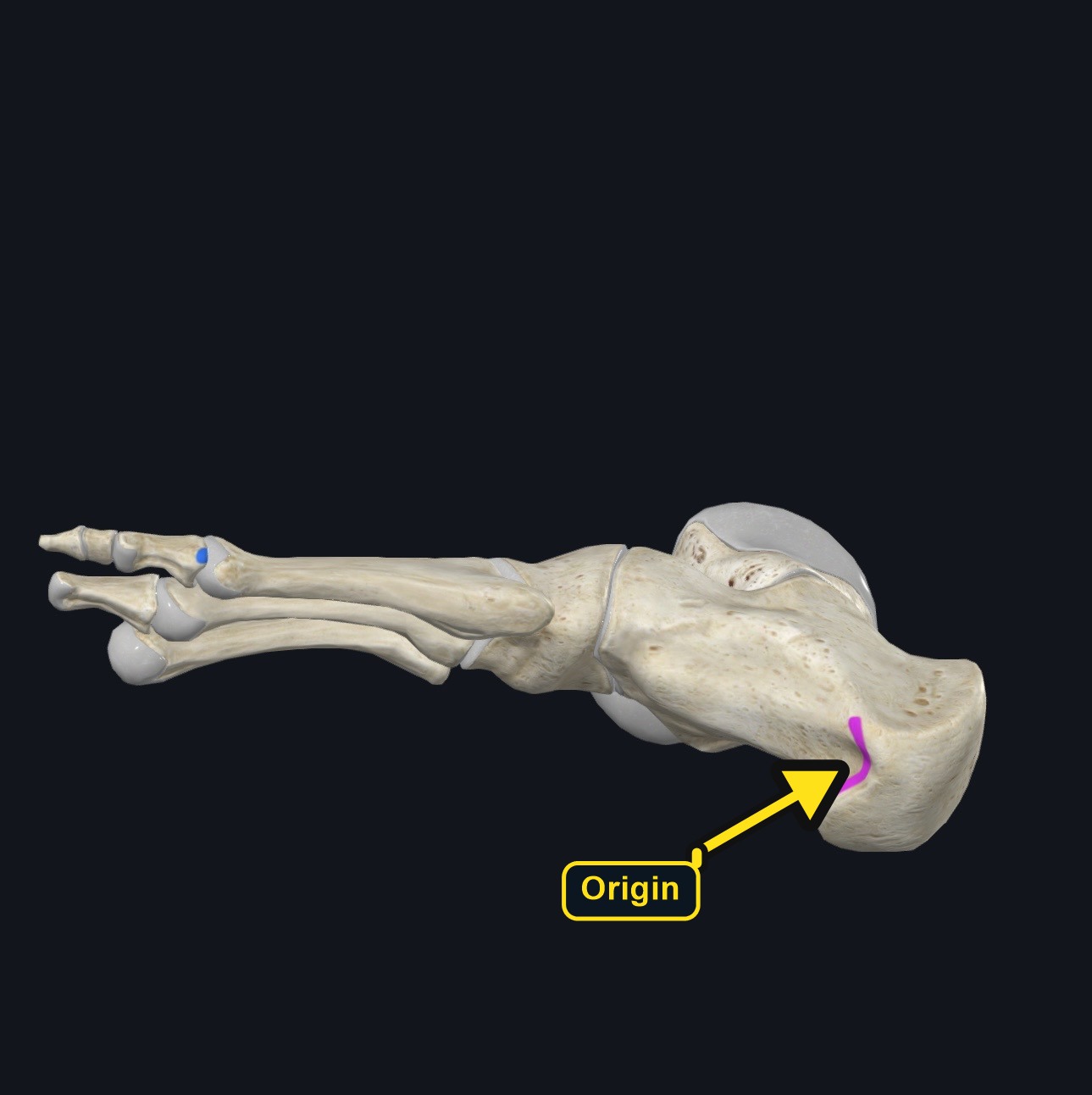
Origin
| Origin | Medial process of the calcaneal tuberosity, the flexor retinaculum, the plantar aponeurosis, and the adjacent intermuscular septum. |
|---|
Section 03 of 20
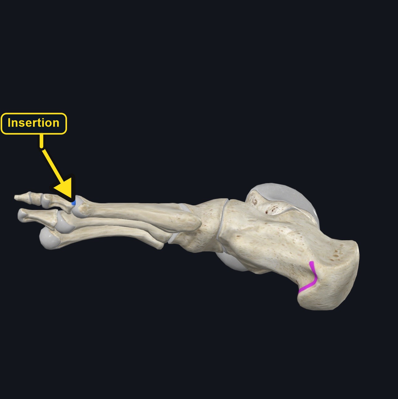
Insertion
| Insertion | Medial side of the base of the proximal phalanx of the great toe, usually together with the medial tendon of flexor hallucis brevis. |
|---|
Section 04 of 20
Actions
No dedicated action video is currently available. This movement is explained below and video will be added when verified source media becomes available.
| Primary action | Abduction of the great toe at the metatarsophalangeal joint. |
|---|---|
| Secondary actions | Assists flexion of the great toe; supports the medial longitudinal arch. |
| Movement at the joint | First metatarsophalangeal joint (abduction, small flexion contribution). |
| Heads / fiber groups | Single muscle belly; no separate heads. |
| Functional examples | Keeping the great toe aligned during push-off in walking and running; spreading the toes; balancing on uneven ground. |
Abducts the great toe
Draws the great toe medially, away from the second toe, at the metatarsophalangeal joint.
Assists great-toe flexion
Works with flexor hallucis brevis to flex the metatarsophalangeal joint.
Supports the medial arch
Helps stabilize the medial longitudinal arch and great-toe alignment during stance and push-off.
Section 05 of 20
Nerve Innervation
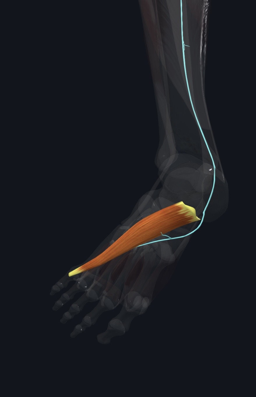
| Nerve | Medial plantar nerve, a branch of the tibial nerve. The segmental level is usually given as S1–S2; sources differ slightly. |
|---|
The medial plantar nerve passes behind the medial malleolus through the tarsal tunnel and then along the abductor hallucis. Medial foot symptoms can therefore reflect the nerve, the muscle, or both, and should not be assumed to be simple muscle tightness.
Section 06 of 20
Blood Supply
| Blood | Medial plantar artery, with contributions from the medial tarsal and first plantar metatarsal branches. |
|---|
Section 07 of 20
Pictures and Visual Anatomy
Section 08 of 20
Functional Movement
During walking and running the muscle helps keep the great toe aligned as the foot rolls onto the forefoot, and it contributes to balance on uneven ground. Weak or overloaded intrinsic foot muscles can change how load is shared across the forefoot.
Section 09 of 20
Synergists and Antagonists
Synergists: flexor hallucis brevis and, for arch support, tibialis posterior and flexor hallucis longus. Antagonists: adductor hallucis, which draws the great toe toward the second toe.
Section 10 of 20
Palpation and Location
Client position and technique
Position the client supine or seated with the foot supported. Ask them to move the great toe gently away from the second toe while you feel along the medial border of the foot, distal to the medial malleolus and below the navicular. The muscle belly is a firm band along the medial arch; its tendon continues toward the base of the great toe.
Protect nearby structures
Use light-to-moderate pressure below and behind the medial malleolus, where the tibial nerve and its plantar branches and the posterior tibial vessels pass. Avoid sustained deep pressure that reproduces tingling, burning, or numbness, and avoid pressing on a tender bunion or inflamed joint.
Section 11 of 20
Massage Therapy Relevance
The foot is a common area of fatigue for people who stand or walk for long periods. Abductor hallucis is a small, superficial muscle that is easy to reach, but medial foot pain has several possible sources, so muscle work should follow a careful history.
Section 12 of 20
Dysfunction: What Happens When Movement Goes Wrong
Changes in how a muscle works can change how a joint moves. The points below describe possibilities that may be associated with altered muscle function; they are not diagnoses, and one muscle is never assumed to be the sole cause of a problem.
When the muscle is weak
Weakness may contribute to poor control of great-toe alignment and can be associated with a flatter or less controlled medial arch, so other foot and calf muscles may work harder during push-off.
When it is tight or shortened
A tight or overworked muscle may be associated with medial arch fatigue and tenderness along the inner border of the foot and may restrict how freely the great toe can move away from the second toe.
When it is overused
Prolonged standing, walking, or unsupportive footwear may load the muscle and may contribute to medial foot ache.
When it is strained
A strain may cause localized pain when the great toe is drawn inward or outward; the pain pattern can resemble other medial foot problems, so it should not be assumed to be muscular.
When it is inhibited or others compensate
When the muscle is inhibited, the great toe may drift toward the second toe and the foot may rely more on the long toe flexors and calf, which can alter gait.
When it does not coordinate well with synergists and antagonists
Poor coordination with flexor hallucis brevis, adductor hallucis, and the arch supporters may change how weight is shared across the forefoot and may be associated with altered push-off.
Section 13 of 20
Conditions, Pathologies, and Pain Relationships
Hallux valgus (bunion)
The muscle can be displaced and altered in long-standing bunions. Massage may support comfort in surrounding tissue but does not correct the joint deformity.
Plantar heel and arch pain
Plantar fasciitis and arch overload can produce medial heel and arch pain near the muscle origin. Sharp first-step heel pain is not automatically an abductor hallucis problem.
Tarsal tunnel or medial plantar nerve irritation
Burning, tingling, or numbness along the medial sole or toes may reflect compression of the tibial nerve branches and needs assessment, not deeper pressure.
Differential considerations
Posterior tibial tendon dysfunction, stress fracture, gout, sesamoid problems, arthritis, peripheral neuropathy, and circulatory disease can all cause medial foot pain.
Section 14 of 20
Example Clinical Relationship
A client who stands all day and reports an aching inner arch may show tenderness along abductor hallucis. This finding may be associated with muscle overload, but medial foot pain can also involve the plantar fascia, the posterior tibial tendon, or the tibial nerve branches. Gentle massage may support comfort, while burning, numbness, or a hot swollen foot needs referral.
Section 15 of 20
Massage and Treatment Approaches
For healthy, non-acute tissue, position the foot in a supported neutral posture and begin with broad effleurage over the sole and medial arch. Progress to gentle compression and short gliding strokes along the muscle belly toward the great toe, keeping pressure comfortable.
| Movement | Use gentle active great-toe abduction and flexion within comfort; do not force stretch into a painful bunion. |
|---|---|
| Regional work | Consider the plantar fascia, calf, tibialis posterior, and other intrinsic foot muscles when assessment supports it. |
| Avoid | Deep pressure over acute pain, a swollen joint, open skin, or the tibial nerve behind the medial malleolus. |
Section 16 of 20
Precautions and Contraindications
Precautions and contraindications
- Acute injury, active inflammation, suspected stress fracture, or recent foot surgery.
- Use caution with diabetes, peripheral neuropathy, poor circulation, or reduced sensation; check skin and use light pressure.
- Avoid open wounds, infection, plantar warts, and tender bunions.
- Ask about anticoagulant use and easy bruising.
Section 17 of 20
Red Flags and When to Refer
Red flags and referral
- Foot or ankle that is red, hot, swollen, and painful, especially with fever.
- Unilateral calf swelling or pain, or sudden shortness of breath.
- Progressive numbness, weakness, or burning that does not settle.
- Cold, pale, or blue toes, non-healing sores, or major trauma.
Section 18 of 20
Study Summary
Attachments
Origin: medial calcaneal tuberosity and plantar aponeurosis. Insertion: medial base of the great-toe proximal phalanx.
Actions
Abducts the great toe, assists flexion, and supports the medial arch.
Supply
Medial plantar nerve (S1–S2) and medial plantar artery.
Section 19 of 20
Instructor Narration
Open narration transcript
Abductor hallucis is the muscle that runs along the inner border of the foot, just below the medial arch. It is the most medial muscle of the first layer of the sole.
It begins on the medial process of the heel bone, the calcaneal tuberosity, with attachments to the flexor retinaculum and plantar aponeurosis. Its tendon inserts on the inner side of the base of the great toe's proximal phalanx, usually blending with the tendon of flexor hallucis brevis.
Its main action is to abduct the great toe, drawing it away from the second toe. It also helps flex the big toe joint and supports the medial longitudinal arch. Its antagonist is adductor hallucis.
The medial plantar nerve, a branch of the tibial nerve, supplies it, usually described as S1 to S2. Blood comes mainly from the medial plantar artery.
To palpate, support the foot and ask the client to move the big toe gently away from the second toe. Feel along the medial border of the foot below the navicular. Stay light behind the medial malleolus, where the tibial nerve and its branches travel.
Bunions, plantar heel pain, tarsal tunnel irritation, posterior tibial tendon problems, and stress fractures can all cause medial foot pain, so muscle tightness should not be assumed. Massage can be broad and gentle in healthy tissue. Refer a hot, red, swollen foot, calf swelling, progressive numbness, cold or blue toes, or a wound that will not heal.
Remember: medial calcaneus to the base of the great toe, abduction of the great toe, medial plantar nerve.
Section 20 of 20